
Page 298 - Read Online
P. 298
Lei et al. HLH presenting as ACS
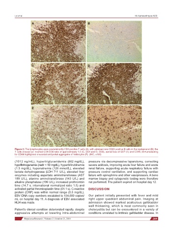
A B
C D
E F
Figure 2: The lymphocytes were predominantly CD3 positive T cells (A), with admixed rare CD20 positive B cells in the background (B); the
T cells showed an inverted CD4:CD8 ratio of approximately 1:2 (C, CD4 and D, CD8), partial loss of CD7 (E) and CD45; immunostaining
for CD68 highlighted sinusoidal and portal aggregates of histiocytes (F). (IHC, ×100)
(1012 ng/mL), hypertriglyceridemia (662 mg/dL), pressure via decompressive laparotomy, correcting
hypofibrinogenemia (nadir < 50 mg/dL), hyperbilirubinemia severe acidosis, improving acute liver failure and acute
(1.3 mg/dL), hyponatremia (126 mmol/L), elevated renal failure, supporting acute respiratory failure with
lactate dehydrogenase (LDH 711 U/L), elevated liver pressure control ventilation, and supporting cardiac
enzymes including aspartate aminotransferase (AST failure with epinephrine and other vasopressors. A bone
189 U/L), alanine aminotransferase (143 U/L) and marrow biopsy and cytogenetic testing were therefore
alkaline phosphatase (196 U/L), increased prothrombin not performed. The patient expired on hospital day 12.
time (14.7 s, international normalized ratio 1.5) and
activated partial thromboplastin time (51.1 s). C-reactive DISCUSSION
protein (CRP) was within normal range (0.5 mg/dL).
EBV DNA copy numbers escalated to 134,000 copies/ Our patient initially presented with fever and mild
mL on hospital day 11. A diagnosis of EBV associated right upper quadrant abdominal pain. Imaging at
HLH was made. admission showed marked acalculous gallbladder
wall thickening, which is most commonly seen in
Patient’s clinical condition deteriorated rapidly, despite cholecystitis but can be encountered in a variety of
aggressive attempts at lowering intra-abdominal conditions unrelated to intrinsic gallbladder disease. In
Hepatoma Research ¦ Volume 2 ¦ October 21, 2016 289