
Page 53 - Read Online
P. 53
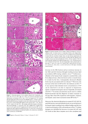
Figure 2: Photomicrographs of liver sections stained with Masson’s trichrome
                                                              staining (×100). (C) Liver section of control rats, fi brosis, and collagen fi bers
                                                              could not be seen; (HCC) liver section of HCC rat treated with thioacetamide,
                                                              increased fi brosis, and blue collagen fi bers are seen among hepatocytes and
                                                              central vein; (HCC + CIS) liver section of HCC rat treated with CIS, strands of blue
                                                              collagen are less seen among hepatocytes; (HCC + IN) liver section of HCC rat
                                                              that received IN, similar to the control rat group; (HCC + CIS + IN) liver section of
                                                              HCC rat treated with both CIS + IN, blue collagen fi bers among hepatocytes are
                                                              less seen. C: control; IN: inulin; CIS: cisplatin; HCC: hepatocellular carcinoma;
                                                              CV: central vein; white arrow: a curved short arrow on the fi gure indicating
                                                              blood sinusoids

                                                              decrease in the HDL fraction and an enormous increase in
                                                              the VLDL and LDL fractions through resulting increase lipid
                                                              peroxidation (LPO) during oxidative stress.  Furthermore,
                                                                                                  [44]
                                                              albumin in the human body transports essential fatty acids
                                                              from adipose tissue, otherwise known as fat, to muscle tissue;
                                                              it is made by the liver. Consequently, decreased albumin
                                                              levels may be associated with liver disease.  Moreover, the
                                                                                                 [45]
                                                              increase in the level of AFP in HCC rats indicates an HCC.
                                                                                                             [46]
                                                              It was reported that elevated serum concentrations of AFP
                                                              can be observed in rats due to exposure to hepatotoxic
                                                              agents or hepatocarcinogens and are frequently associated
                                                              with HCC.  Its serum concentration can be used to confirm
                                                                      [47]
                                                              hepatocarcinoma and the diagnosis of tumor response to
          Figure 1: Photomicrographs of liver sections stained with hematoxylin and   therapy. More than 90% of patients with hepatitis, cirrhosis,
          eosin (HE, left row ×100, right row ×400). (C) Control rat showing normal   and hepatic cancer have increased serum AFP levels. [47]
          hepatocytes architecture; (HCC) liver of HCC rat treated with CIS, showing
          damage hepatocytes with lymphocyte cells infi ltration; (HCC + CIS) liver of HCC rat
          treated with CIS showing congested central vein and blood sinusoids; (HCC + IN)   Moreover, the observed alterations in serum AST, ALT, ALP, CK,
          liver of HCC rat that received IN similar to control group; (HCC + CIS + IN) liver   and LDH activity and total bilirubin levels are good indicators
          of HCC treated with both CIS + IN showing maintained hepatic architecture,
          with minimal damage. C: control; IN: inulin; CIS: cisplatin; HCC: hepatocellular   for hepatic injury resulting from cellular leakage and the loss
          carcinoma; CV: central vein; HP: hepatocytes; BS: blood sinusoids; VHP: vascular   in functional integrity of the cell membrane in liver.  Cellular
                                                                                                       [48]
          hepatocytes; DBS: degenerated blood sinusoids; PV: portal vein; H: hepatocytes;   damages are identified by increases in serum ALP, ALT, and
          white arrow: blood sinusoids; arrowhead: Kupffer cells; black arrow: endothelial
          cells; *Lymphocytic cell infi ltration               AST levels in that these enzymes are in the cytoplasm and

               Hepatoma Research | Volume 1 | Issue 3 | October 15, 2015                                    151