
Page 52 - Read Online
P. 52
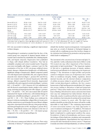
Table 2: Serum and liver enzyme activity in control and treated rat groups
          Parameters                                             Animal groups
                                Control            TAA            TAA + CIS         TAA + IN         TAA + IN +
                                                                                                        CIS
          Serum AST (U/L)     35.00 ± 3.20 a   200.76 ± 3.33 b   123.06 ± 1.19 c   118.81 ± 2.35   c  86.96 ± 2.23 d
          Serum ALT (U/L)     56.53 ± 1.85 a   199.00 ± 2.89 b   96.16 ± 3.50 c    77.08 ± 1.80 d    60.53 ± 2.62 a
          Serum ALP (U/L)     6.44 ± 0.30 a     11.77 ± 0.33 b    8.95 ± 0.24 c     8.14 ± 0.19 c    7.32 ± 0.39 d
          Serum CK (U/L)      241.16 ± 0.97 a  482.81 ± 7.05 b   308.91 ± 4.39 c  278.28 ± 2.72 d   259.96 ± 1.04 e
          Serum              218.33 ± 10.77 a  468.33 ± 9.36 b  297.83 ± 16.03 c   291.33 ± 0.18 c  279.50 ± 10.44 c
          LDH (U/L)
          Liver AST (U/g)      8.94 ± 0.16 a    0.09 ± 0.42 b     2.21 ± 0.38 c     3.96 ± 0.15 d    5.43 ± 0.23 e
          Liver ALT (U/g)     4.65 ± 0.31 a     0.63 ± 0.06 b     1.55 ± 0.15 c     1.93 ± 0.12 c    2.68 ± 0.35 d
          Liver ALP (U/g)     50.97 ± 1.93 a   23.19 ± 1.43 b    36.11 ± 1.29 c    39.76 ± 0.51 c    48.35 ± 1.41 a
          Results are expressed as a mean ± SEM, with each row. Superscripts with different letters (a-e) express signifi cant change with P ≤ 0.05. Superscripts with
          similar letters were non-signifi cant. Means with different letters were signifi cant (P ≤ 0.05, n = 6). TAA: thioacetamide; IN: inulin; CIS: cisplatin; AST: aspartate
          transaminase; ALT: alanine transaminase; ALP: alkaline phosphatase; CK: creatine kinase; LDH: lactate dehydrogenase; SEM: standard error of mean

          HCC rats succeeded in inducing a significant improvement   stimuli that mediate hepatocarcinogenesis. Carcinogenesis
          in these changes.                                   may arise as a result of chemical or biological damage to
                                                              normal cells in a multistep process that involves changes at
          Histopathological examination revealed that the liver of the   the initiation level followed by promotion and progression,
          control group was composed of classical hepatic lobules of   which leads to malignancy. [37]
          normal central veins lined by endothelial cells, radiating hepatic
          cells, and hepatic sinusoids. Hepatocytes were polyhedral   The increment in the concentrations of serum total lipid, TC,
          in shape, with sharply defined boundaries. They had an   TGs, and LDL-C with a reduction in the level of HDL-C in the
          acidophilic cytoplasm and central rounded nuclei. Frequently   TAA treated rats may reflect impairment of liver function,
          seen were von Kupffer cells [Figure 1, pictures C and C1]. The   particularly in lipid metabolism.  The long-term regimen
                                                                                         [38]
          examination of the liver in HCC rat groups exhibited severe   of TAA led to a significant decrease of hepatic markers;
          hepatic damages. Inflammation (lymphocytic cell infiltration),   total protein and albumin levels indicating acute hepatocyte
          significant damage of hepatocytes, dilated congested central   damage.  The alterations in lipid profiles and protein
                                                                      [39]
          vein with degenerated endothelial cells, and congested blood   content in malignant tissues are of importance due to their
          sinusoids were observed [Figure 1, pictures HCC and HCC1].   effect on membrane integrity, fluidity, regulation, altered
          HCC rats treated with CIS showed hepatocytes with little   internal viscosity, and the internal chemical composition
          damage, dilated congested blood sinusoids, degenerated   of cellular processes related to growth and cell survival.
                                                                                                             [40]
          endothelial cells, and congested central veins [Figure 1, pictures   Under normal physiological conditions, the liver ensures
          HCC + CIS and HCC + CIS1]. However, HCC rats administered   homeostasis of lipid and lipoprotein metabolism. HCC impairs
          with IN showed normal hepatic lobules similar to the control   this process, leading to alterations in lipid and lipoprotein
          group, indicating its hepatoprotective effect [Figure 1, pictures   patterns.  Many tumor markers have been described in the
                                                                      [41]
          HCC + IN and HCC + IN1]. HCC rats treated with CIS and IN   hope of finding a blood test for cancer, and some have found
          maintained the hepatic architecture, with minimal damage in   their way into widespread but indiscriminate clinical use.
          lymphocytic cell infiltration [Figure 1, pictures HCC + CIS + IN   Classically, a marker is synthesized by the tumor and released
          and HCC + CIS + IN1]. These findings were evidenced by   into circulation, but it may also be produced by normal tissue
          Masson’s trichrome staining blue collagen fibers in hepatocytes   in response to invasion by cancer cells. The ideal tumor
          and the central vein as a good marker for more fibrous collagen   markers should be produced by the tumor cell and be readily
          deposition, developed extensive fibrosis in the periportal area,   detectable in body fluids.  The continuing improvement
                                                                                    [42]
          and damaged hepatocytes in HCC rats [Figure 2, picture HCC].   of tumor-associated markers may help approaches in
          Whereas, small fibrotic lesions were detected in the liver of   cancer treatment and diagnosis.  In addition, TAA-induced
                                                                                        [42]
          HCC + IN, HCC + CIS and HCC + IN + CIS treated groups   abnormal lipid synthesis or defective degradation of
          compared to the control rat group [Figure 2].       lipids is implicated in a pathological condition like cancer.
                                                              Peroxidation of lipids in biomembranes and tissues causes
          DISCUSSION                                          the leakage of these lipids into circulation and consequently
                                                              leads to hyperlipidemia. Hyperlipidemia has been shown to
          HCC is the most common and lethal of all cancers. There exists   increase the risk of metastasis in several cancers.  Hepatoma
                                                                                                     [43]
          a diversity of dietary,  endogenous, and environmental    is usually associated with hyperlipidemia, as well as a notable
                                                         [36]
                            [35]
          150                                                       Hepatoma Research | Volume 1 | Issue 3 | October 15, 2015