
Page 51 - Read Online
P. 51
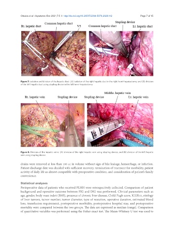
Otsuka et al. Hepatoma Res 2021;7:5  I  http://dx.doi.org/10.20517/2394-5079.2020.112                                        Page 7 of 15


                 A                                             B


               Figure 7. Isolation and division of the hepatic duct: (A) isolation of the right hepatic duct in the right hemi-hepatectomy; and (B) division
               of the left hepatic duct using stapling device in the left hemi-hepatectomy


                A                                             B


               Figure 8. Division of the hepatic veins: (A) division of the right hepatic vein using stapling device; and (B) division of the left hepatic
               vein using stapling device

               drains were removed at less than 100 cc in volume without sign of bile leakage, hemorrhage, or infection.
               Patient discharge date was decided with sufficient recovery, termination of treatment for morbidity, patient
               activity of daily life as almost compatible with preoperative condition, and consideration of patient’s family
               convenience.


               Statistical analyses
               Perioperative data of patients who received PLHH were retrospectively collected. Comparison of patient
               background and operative outcome between FIG and DIG was performed. Clinical parameters such as
               age, gender, body mass index (BMI), presence of chronic liver disease, Child-Pugh score, ICGR15, etiology
               of liver tumors, tumor number, tumor diameter, type of resection, operative duration, estimated blood
               loss, transfusion requirement, postoperative morbidity, postoperative hospital stay, and postoperative
               mortality were compared between the two groups. The data are expressed as median (range). Comparison
               of quantitative variables was performed using the Fisher exact test. The Mann-Whitney U test was used to