
Page 54 - Read Online
P. 54
Page 10 of 15 Otsuka et al. Hepatoma Res 2021;7:5 I http://dx.doi.org/10.20517/2394-5079.2020.112
Table 3. Comparison of operative outcome of pure laparoscopic hemi-hepatectomy
Favorable cases (n = 11) Difficult cases (n = 10) P-value
Operative duration 586.0 (355-749) 625.5 (240-768) 0.251
Estimated blood loss 290.0 (10-1060) 357.5 (50-2683) 0.342
Transfusion requirement 1 (9.1%) 2 (20.0%) 0.586
Postoperative morbidity 2 (18.2%) 3 (30.0%) 0.635
SSI (organ/space) 1 (9.1%) SSI (superficial) 1(10.0%)
Bile leakage 1 (9.1%) Bile leakage 1(10.0%)
Portal vein thrombus 1(10.0%)
Postoperative hospitalization 9(6-25) 13(7-45) 0.111
Mortality 0 (0.0%) 0 (0.0%) 1.0
A B
C D
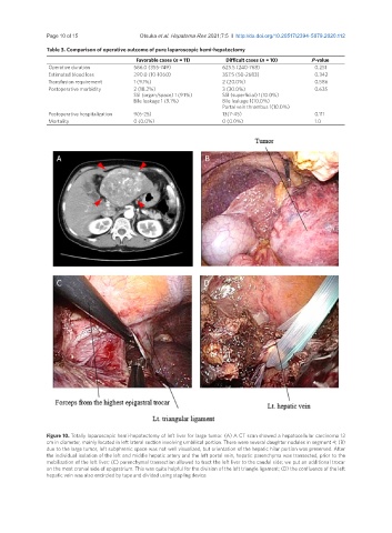
Figure 10. Totally laparoscopic hemi-hepatectomy of left liver for large tumor. (A) A CT scan showed a hepatocellular carcinoma 12
cm in diameter, mainly located in left lateral section involving umbilical portion. There were several daughter nodules in segment 4; (B)
due to the large tumor, left subphrenic space was not well visualized, but orientation of the hepatic hilar portion was preserved. After
the individual isolation of the left and middle hepatic artery and the left portal vein, hepatic parenchyma was transected, prior to the
mobilization of the left liver; (C) parenchymal transection allowed to tract the left liver to the caudal side; we put an additional trocar
on the most cranial side of epigastrium. This was quite helpful for the division of the left triangle ligament; (D) the confluence of the left
hepatic vein was also encircled by tape and divided using stapling device