
Page 53 - Read Online
P. 53
Otsuka et al. Hepatoma Res 2021;7:5 I http://dx.doi.org/10.20517/2394-5079.2020.112 Page 9 of 15
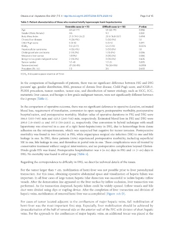
Table 2. Patient characteristics of those who received totally laparoscopic hemi-hepatectomies
Favorable cases (n = 11) Difficult cases (n = 10) P-value
Age 60 (41-77) 53 (26-79) 0.382
Gender (Male: Female) 4:6 9:2 0.361
Body Mass Index 21.3 (19.0-26.2) 23.5 (16.8-35.1) 0.494
Chronic liver disease 4 (36.4%) 5 (50.0%) 1.0
Child-Pugh score 5 (5-8) 5 (5-5) 0.391
9.0 (2-17) 5.5 (1-10) 0.0576
ICGR 15
Hepatocellular carcinoma 4 (36.4%) 5 (50.0%) 1.0
Cholangiocellular carcinoma 2 (18.2%) 1 (10.0%) 0.586
Metastatic liver cancer 1 (9.1%) 3 (30.0%) 0.586
Benign to low grade malignant tumor 2 (18.2%) 3 (30.0%) 0.635
Tumor-number 1 (1-4) 1 (1-5) 0.895
Tumor-size(mm) 47 (30-55) 70 (33-120) 0.0559
Procedure (Rt.: Lt.) 7:3 3:5 0.659
ICGR 15 : Indocyanine green retention at 15min
In the comparison of backgrounds of patients, there was no significant difference between FIG and DIG
patients’ age, gender distribution, BMI, presence of chronic liver disease, Child-Pugh score, and ICGR15.
PLHH procedure, tumor-number, tumor-size, and distribution of tumor etiology, such as HCC, ICC,
metastatic liver cancer, and benign to low grade malignant tumors, were not significantly different between
the 2 groups [Table 2].
In the comparison of operative outcome, there was no significant deference in operative duration, estimated
blood loss, requirement of transfusion, conversion to open surgery, postoperative morbidity, postoperative
hospitalization, and postoperative mortality. Median value of operative durations in FIG and DIG were
586.0 (355-749) min and 625.5 (240-768) min, respectively. Estimated blood loss in FIG and DIG were
290.0 (10-1060) cc and 357.5 (50-2683) cc, respectively. One conversion to hybrid technique with small
laparotomy was observed in a case of a right hemi-hepatectomy in DIG, due to hemorrhage from dense
adhesion on the retroperitoneum, which was suspected but negative for tumor invasion. Postoperative
morbidity was found in two (18.2%) in FIG, while organ/space surgical site infection (SSI) in one and bile
leakage in one. In DIG, three patients (30%) experienced postoperative morbidity, including superficial
SSI in one, bile leakage in one, and thrombus in portal vein in one. These complications were all treated by
conservative treatment without surgical interventions, and no postoperative complication beyond Clavien-
Dindo grade IIIb was found. Postoperative hospitalization was 9 (6-25) days in FIG and 13 (7-45) days in
DIG. No mortality was found in either group [Table 3].
Regarding the correspondence to difficulty in DIG, we describe technical details of the issues.
For the tumor larger than 7 cm, mobilization of hemi-liver was not possible prior to liver parenchymal
transection. For this issue, obtaining operative abdominal space and visualization of hepatic hilum was
important. In all four cases of large tumor, hepatic hilar dissection was successful to isolate hepatic inflow
vessels. After the demarcation line appeared on the liver surface by inflow occlusion, liver transection was
performed. As the transection deepened, hepatic hilum could be widely opened. Inflow vessels and bile
duct were divided using clips or stapling device. After the completion of liver transection and division of
hepatic veins, mobilization of removed hemi-liver was accomplished [Figure 10A-D].
For cases of tumor located adjacent to the confluences of major hepatic veins, full mobilization of
hemi-liver was the most important first step. Especially, liver mobilization should be achieved by
devascularization of the half of removal side on the anterior wall of the IVC with division of short hepatic
veins. For the approach to the confluences of major hepatic veins, an additional trocar was placed at the