
Page 48 - Read Online
P. 48
Page 4 of 15 Otsuka et al. Hepatoma Res 2021;7:5 I http://dx.doi.org/10.20517/2394-5079.2020.112
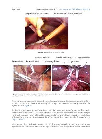
Figure 2. Extra corporeal Pringle’s tourniquet
A B
Figure 3. Dissection of hepatic hilum using individual isolation approach: (A) hepatic hilar dissection in the right hemi-hepatectomy;
and (B) hepatic hilar dissection in the left hemi-hepatectomy
After conventional laparoscopic cholecystectomy, the hepatoduodenal ligament was encircled by tape.
Furthermore, an extracorporeal Rumel tourniquet for Pringle’s maneuver was made using catheter on left
hypochondrium [Figure 2].
For hepatic inflow control, we usually performed individual isolation technique for hepatic inflow vessels.
The hepatic hilar dissection was performed. The anterior and posterior branch of the right hepatic artery in
right hemi-hepatectomy and the left and the middle hepatic artery in left hemi-hepatectomy were isolated
and taped. With retraction of these arteries, the right or left portal vein was dissected and isolated by tape
[Figure 3A and B].
These hepatic inflow vessels were temporarily occluded individually in order to confirm the ischemic area
appeared on the liver surface. After that, the hepatic artery was doubly clipped and divided. The right or