
Page 47 - Read Online
P. 47
Otsuka et al. Hepatoma Res 2021;7:5 I http://dx.doi.org/10.20517/2394-5079.2020.112 Page 3 of 15
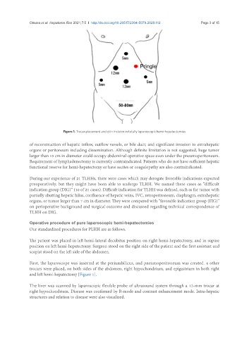
Figure 1. Trocar placement and skin incision in totally laparoscopic hemi-hepatectomies
of reconstruction of hepatic inflow, outflow vessels, or bile duct; and significant invasion to extrahepatic
organs or peritoneum including dissemination. Although definite limitation is not suggested, huge tumor
larger than 15 cm in diameter could occupy abdominal operative space even under the pneumoperitoneum.
Requirement of lymphadenectomy is currently contraindicated. Patients who do not have sufficient hepatic
functional reserve for hemi-hepatectomy or have ascites or coagulopathy are also contraindicated.
During our experience of 21 TLHHs, there were cases which may derogate favorable indications expected
preoperatively, but they might have been able to undergo TLHH. We named these cases as “difficult
indication group (DIG)” (10 of 21 cases). Difficult indication for TLHH was defined, such as for tumor with
partially abutting hepatic hilus, confluence of hepatic veins, IVC, retroperitoneum, diaphragm, extrahepatic
organs, or tumor larger than 7 cm in diameter. They were compared with “favorable indication group (FIG)”
on perioperative background and surgical outcome and discussed regarding technical correspondence of
TLHH on DIG.
Operative procedure of pure laparoscopic hemi-hepatectomies
Our standardized procedures for PLHH are as follows.
The patient was placed in left hemi-lateral decubitus position on right hemi-hepatectomy, and in supine
position on left hemi-hepatectomy. Surgeon stood on the right side of the patient and the first assistant and
scopist stood on the left side of the abdomen.
First, the laparoscope was inserted at the periumbilicus, and pneumoperitoneum was created. 4 other
trocars were placed, on both sides of the abdomen, right hypochondrium, and epigastrium in both right
and left hemi-hepatectomy [Figure 1].
The liver was scanned by laparoscopic flexible probe of ultrasound system through a 12-mm trocar at
right hypochondrium. Disease was confirmed by B-mode and contrast enhancement mode. Intra-hepatic
structures and relation to disease were also visualized.