
Page 49 - Read Online
P. 49
Otsuka et al. Hepatoma Res 2021;7:5 I http://dx.doi.org/10.20517/2394-5079.2020.112 Page 5 of 15
A B
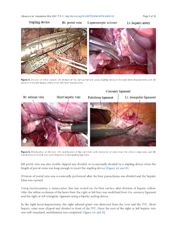
Figure 4. Division of inflow vessels: (A) division of the right portal vein using stapling device in the right hemi-hepatectomy; and (B)
division of the left hepatic artery in the left hemi-hepatectomy
A B
Figure 5. Mobilization of the liver: (A) mobilization of the right liver with dissection of veins from the inferior vena cava; and (B)
mobilization of the left liver with dissection of surrounding ligaments
left portal vein was also doubly clipped and divided, or occasionally divided by a stapling device when the
length of portal veins was long enough to insert the stapling device [Figure 4A and B].
Division of portal vein was occasionally performed after the liver parenchyma was divided and the hepatic
hilus was opened.
Using electrocautery, a demarcation line was scored on the liver surface after division of hepatic inflow.
After the inflow occlusion of the hemi-liver, the right or left liver was mobilized from the coronary ligament
and the right or left triangular ligament using a bipolar sealing device.
In the right hemi-hepatectomy, the right adrenal gland was dissected from the liver and the IVC. Short
hepatic veins were clipped and divided in front of the IVC. Once the root of the right or left hepatic vein
was well visualized, mobilization was completed [Figure 5A and B].