
Page 55 - Read Online
P. 55
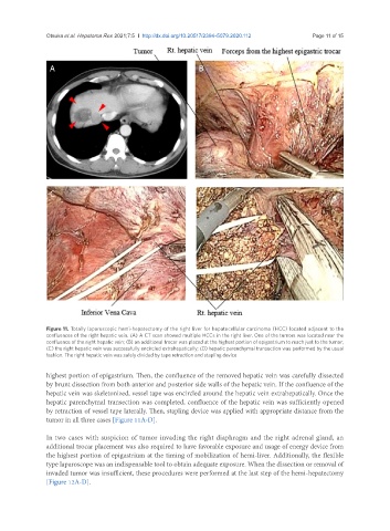
Otsuka et al. Hepatoma Res 2021;7:5  I  http://dx.doi.org/10.20517/2394-5079.2020.112                                        Page 11 of 15


                A                                             B


                C                                             D


               Figure 11. Totally laparoscopic hemi-hepatectomy of the right liver for hepatocellular carcinoma (HCC) located adjacent to the
               confluences of the right hepatic vein. (A) A CT scan showed multiple HCCs in the right liver. One of the tumors was located near the
               confluence of the right hepatic vein; (B) an additional trocar was placed at the highest portion of epigastrium to reach just to the tumor;
               (C) the right hepatic vein was successfully encircled extrahepatically; (D) hepatic parenchymal transection was performed by the usual
               fashion. The right hepatic vein was safely divided by tape retraction and stapling device

               highest portion of epigastrium. Then, the confluence of the removed hepatic vein was carefully dissected
               by brunt dissection from both anterior and posterior side walls of the hepatic vein. If the confluence of the
               hepatic vein was skeletonized, vessel tape was encircled around the hepatic vein extrahepatically. Once the
               hepatic parenchymal transection was completed, confluence of the hepatic vein was sufficiently opened
               by retraction of vessel tape laterally. Then, stapling device was applied with appropriate distance from the
               tumor in all three cases [Figure 11A-D].

               In two cases with suspicion of tumor invading the right diaphragm and the right adrenal gland, an
               additional trocar placement was also required to have favorable exposure and usage of energy device from
               the highest portion of epigastrium at the timing of mobilization of hemi-liver. Additionally, the flexible
               type laparoscope was an indispensable tool to obtain adequate exposure. When the dissection or removal of
               invaded tumor was insufficient, these procedures were performed at the last step of the hemi-hepatectomy
               [Figure 12A-D].