
Page 33 - Read Online
P. 33
Page 8 of 14 Kobayashi et al. Hepatoma Res 2020;6:36 I http://dx.doi.org/10.20517/2394-5079.2020.24
A B
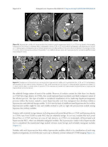
Figure 10. Hypovascular nodule with hypervascular foci (nodule in nodule appearance lesion). A: on CT during hepatic arteriography,
hypervascular foci (arrow) is observed within hypovascular nodule (*); B: on CT during arterial portography, only hypervascular portion
on CT during hepatic arteriography (arrow) shows hypointensity and other portion of the nodule show isointensity to the background liver
(*). In this nodule hypovascular portion of the nodule (*) are relatively benign nature and hypervascular foci represents dedifferentiated
more malignant nature
A B C
Figure 11. Development of hepatocellular carcinoma from hypovascular nodule with hypervascular foci. A, B: on CT during hepatic
arteriography (A) and arterial phase of dynamic CT (B), small hypervascular foci (arrow) is observed within hypovascular nodule (*);
C: eleven months after, on arterial phase of dynamic CT, the hepovascular nodule with hypervascular foci transformed to hypervascular
hepatocellular carcinoma (arrow)
the relatively benign nature of most of the nodule. However, if nodules contain foci that show low density
on CTAP but a high density on CTHA, this would represent hypervascularity and likely malignant nature of
the affected portion. This type of nodule is considered visualization of the multi-step hepatocarcinogenesis
process within the tumor, namely a more hypervascular and thus malignant foci develops within a
hypovascular and relatively benign nodule. To find out this kind of dedifferentiated hypervascular foci within
hypovascular nodule might be easy and reliable method to pick up high-risk precancerous nodule among
hypovascular cirrhotic nodules.
Nodules with relatively benign features including preserved portal blood flow on CTAP and hypovascularity
on CTHA vary from LGDN to early HCC that are relatively benign. In contrast, nodules that lack portal
blood flow on CTAP and have an area of high density on CTHA are moderately differentiated and
hypervascular HCC, which are definitely malignant nodules. The hypovascular nodule with a hypervascular
foci is considered the transitional stage nodule between more benign and definitely malignant nodules
[Figure 11].
Nodules with such hypervascular focis within hypovascular nodules, which is the visualization of multi-step
hepatocarcinogenesis, are relatively easy to pick up on dynamic contrast enhanced CT/MR imaging [Figure 12],