
Page 32 - Read Online
P. 32
Kobayashi et al. Hepatoma Res 2020;6:36 I http://dx.doi.org/10.20517/2394-5079.2020.24 Page 7 of 14
Figure 8. Multistep changes of drainage vessels and peritumoral enhancement during hepatocarcinogenesis. HCC: hepatocellular
carcinoma
A B
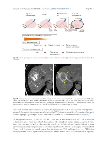
Figure 9. Example of corona enhancement caused by drainage flow from hypervascular HCC. A: on early phase image of CT during
hepatic arteriography, HCC shows marked enhancement because of hypervascularity (*); B: on late phase image of CT during hepatic
arteriography, since the intraarterial contrast injection is stopped, the enhancement of HCC decreases (*) and peritumoral mormal liver
parenchyma is enhanced by drainage of contrast material from HCC (arrow). HCC: hepatocellular carcinoma
connection between tumor sinusoids and surrounding hepatic sinusoids is lost, such that drainage flow is
retrograde through the intracapsular portal venules. On CTHA, this retrograde flow of contrast medium stains
[11]
surrounding hepatic parenchyma around the tumor and is identified as corona enhancement [Figure 9] .
On angiography-assisted CT, LGDN, early HCC, and part of well-differentiated HCC are all observed
as hypovascular nodules. In contrast, the presence of a nodule-in-nodule appearance, which has a
partly hypervascular foci within a hypovascular nodule, is a definite hallmark to discriminate between
hypovascular, relatively benign nodules and more malignant nodules since the latter display hypervascularity
[Figure 10]. In hypovascular nodules, most show iso-density on CTAP and hypo-density on CTHA since
nodular portal blood flow is preserved and increase in hepatic arterial flow has not started, which represents