
Page 28 - Read Online
P. 28
Kobayashi et al. Hepatoma Res 2020;6:36 I http://dx.doi.org/10.20517/2394-5079.2020.24 Page 3 of 14
Figure 1. Schematic diagram of multistep hepatocarcinogenesis. Two types of human hepatocarcinogenesis are currently considered, one
is multistep- and the other is de novo-hepatocarcinogenesis. In multistep hepatocarcinogenesis, the nodular grade of malignancy changes
from dysplastic nodule to advanced HCC stepwisely, and nodular vascularity also gradually change from hypovascular to hypervascular.
HCC: hepatocellular carcinoma
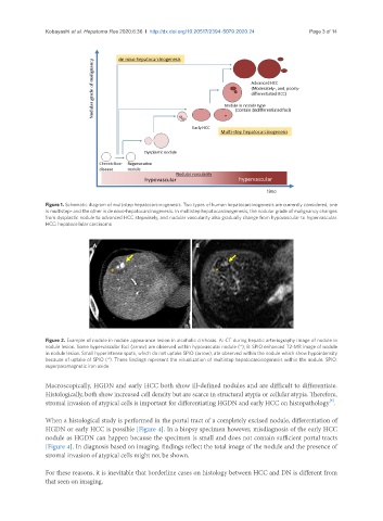
A B
Figure 2. Example of nodule in nodule appearance lesion in alcoholic cirrhosis. A: CT during hepatic arteriography image of nodule in
nodule lesion. Some hypervascular foci (arrow) are observed within hypovascular nodule (*); B: SPIO enhanced T2-MR image of nodule
in nodule lesion. Small hyperintense spots, which do not uptake SPIO (arrow), ate observed within the nodule which show hypointensity
because of uptake of SPIO (*). These findings represent the visualization of multistep hepatocarcinogenesis within the nodule. SPIO:
superparamagnetic iron oxide
Macroscopically, HGDN and early HCC both show ill-defined nodules and are difficult to differentiate.
Histologically, both show increased cell density but are scarce in structural atypia or cellular atypia. Therefore,
[5]
stromal invasion of atypical cells is important for differentiating HGDN and early HCC on histopathology .
When a histological study is performed in the portal tract of a completely excised nodule, differentiation of
HGDN or early HCC is possible [Figure 4]. In a biopsy specimen however, misdiagnosis of the early HCC
nodule as HGDN can happen because the specimen is small and does not contain sufficient portal tracts
[Figure 4]. In diagnosis based on imaging, findings reflect the total image of the nodule and the presence of
stromal invasion of atypical cells might not be shown.
For these reasons, it is inevitable that borderline cases on histology between HCC and DN is different from
that seen on imaging.