
Page 30 - Read Online
P. 30
Kobayashi et al. Hepatoma Res 2020;6:36 I http://dx.doi.org/10.20517/2394-5079.2020.24 Page 5 of 14
A B
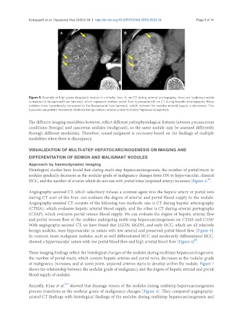
Figure 5. Example of high grade dysplastic nodule in cirrhotic liver. A: on CT during arterial portography, there are isodense nodule
compared to background liver (arrows), which represent nodular portal flow is preserved; B: on CT during hepatic arteriography, these
nodules show hypodensity compared to the background liver (arrows), which indicate the nodular arterial supply is decreased. This
hypovascular pattern represents relatively benign nature nodules under multistep hepatocarcinogenesis
The different imaging modalities however, reflect different pathophysiological features between precancerous
conditions (benign) and cancerous nodules (malignant), so the same nodule may be assessed differently
through different modalities. Therefore, sound judgment is necessary based on the findings of multiple
modalities when there is discrepancy.
VISUALIZATION OF MULTI-STEP HEPATOCARCINOGENESIS ON IMAGING AND
DIFFERENTIATION OF BENIGN AND MALIGNANT NODULES
Approach by haemodynamic imaging
Histological studies have found that during multi-step hepatocarcinogenesis, the number of portal tracts in
nodules gradually decreases as the nodular grade of malignancy changes from DN to hypervascular, classical
[8]
HCC, and the number of arteries which do not run with portal veins (unpaired artery) increases [Figure 3] .
Angiography-assisted CT, which selectively infuses a contrast agent into the hepatic artery or portal vein
during CT scan of the liver, can evaluate the degree of arterial and portal blood supply to the nodule.
Angiography-assisted CT consists of the following two methods: one is CT during hepatic arteriography
(CTHA), which evaluates hepatic arterial blood supply, and the other is CT during arterial portography
(CTAP), which evaluates portal venous blood supply. We can evaluate the degree of hepatic arterial flow
and portal venous flow of the nodules undergoing multi-step hepatocarcinogenesis on CTHA and CTAP.
With angiography-assisted CT, we have found that LGDN, HGDN, and early HCC, which are all relatively
benign nodules, were hypovascular in nature with low arterial and preserved portal blood flow [Figure 5].
In contrast, more malignant nodules, such as well differentiated HCC and moderately differentiated HCC,
[9]
showed a hypervascular nature with low portal blood flow and high arterial blood flow [Figure 6] .
These imaging findings reflect the histological changes of the nodules during multistep hepatocarcinogenesis:
the number of portal tracts, which contain hepatic arteries and portal veins, decreases as the nodular grade
of malignancy increases, and at some point, unpaired arteries starts to develop within the nodule. Figure 7
shows the relationship between the nodular grade of malignancy and the degree of hepatic arterial and portal
blood supply of nodules.
[10]
Recently, Kitao et al. showed that drainage vessels of the nodules during multistep hepatocarcinogenesis
process transform as the nodular grade of malignancy changes [Figure 8]. They compared angiography-
asisted CT findings with histological findings of the nodules during multistep hepatocarcinogenesis and