
Page 35 - Read Online
P. 35
Page 10 of 14 Kobayashi et al. Hepatoma Res 2020;6:36 I http://dx.doi.org/10.20517/2394-5079.2020.24
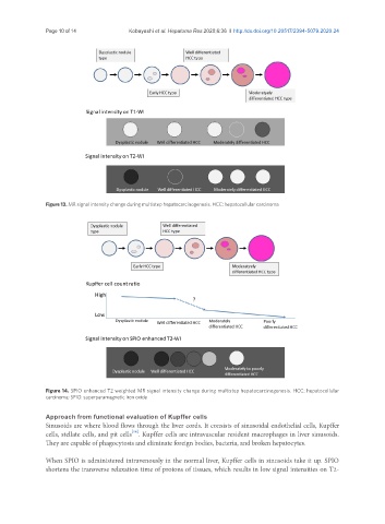
Figure 13. MR signal intensity change during multistep hepatocarcinogenesis. HCC: hepatocellular carcinoma
Figure 14. SPIO enhanced T2 weighted MR signal intensity change during multistep hepatocarcinogenesis. HCC: hepatocellular
carcinoma; SPIO: superparamagnetic iron oxide
Approach from functional evaluation of Kupffer cells
Sinusoids are where blood flows through the liver cords. It consists of sinusoidal endothelial cells, Kupffer
[16]
cells, stellate cells, and pit cells . Kupffer cells are intravascular resident macrophages in liver sinusoids.
They are capable of phagocytosis and eliminate foreign bodies, bacteria, and broken hepatocytes.
When SPIO is administered intravenously in the normal liver, Kupffer cells in sinusoids take it up. SPIO
shortens the transverse relaxation time of protons of tissues, which results in low signal intensities on T2-