
Page 37 - Read Online
P. 37
Page 12 of 14 Kobayashi et al. Hepatoma Res 2020;6:36 I http://dx.doi.org/10.20517/2394-5079.2020.24
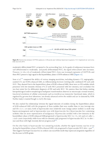
Figure 15. Relationship between OATP1B3 expression, EOB uptake and multistep hepatocarcinogenesis. HCC: hepatocellular carcinoma;
EOB: Gd-EOB-DTPA
moderately differentiated HCC compared to the surrounding liver. As the grade of malignancy increases from
well-differentiated to moderately- and poorly-differentiated HCC, the signal enhancement ratio decreases.
However, in 10%-15% of moderately differentiated HCC, there was increased expression of OATP1B3 and
these HCC present a high signal in the hepatobiliary phase of EOB-enhanced MRI [Figure 15].
[21]
Sano et al. compared the ability of various imaging modalities, including dynamic CT, angiography-
assisted CT, and EOB-enhanced MRI, in differentiating between histologically confirmed DN and early
HCC. They showed that in the hepatobiliary phase of EOB-enhanced MRI, there were no DN nodules which
presented with low-intensity whereas 97% of early HCC presented with low-intensity, and concluded this
was best suited for the differential diagnosis of DN and early HCC. We surmise then that before starting
histopathological and/or morphological malignant transformation observed as microscopic stromal invasion,
functional alterations of cellular components such as membranous transporter functional states, which is
regulated by genome level alterations, are already advanced within the nodule and can be detected by EOB.
Further study is needed though to clarify this issue.
We also studied the relationship between the signal intensity of nodules during the hepatobiliary phase
of EOB-enhanced MRI with the prognosis of these nodules that were smaller than 20 mm (average size
and SD; 11.0 ± 2.8 mm), both in hypovascular ones (relatively more benign nature nodule) in multi-step
hepatocarcinogenesis and in hypovascular ones with hypervascular foci (hypervascular foci-containing
nodules) that are more representative of malignancy. Hypovascular nodules with a low-intensity in the
hepatobiliary phase of EOB-enhanced MRI,progressed to hypervascular HCC in 17%, 28% and 41% after 1,
2 and 3 years respectively, while those with iso-intensity only progressed to hypervascular HCC in 7% after 1
[22]
year and those with high-intensity did not progress at all .
On the other hand, in hypervascular foci-containing nodules, whether the signal intensity was low or iso
[23]
during the hepatobiliary phase of EOB-enhanced MRI, 50% progressed to hypervascular HCC after 1 year .
This suggests that hypervascular foci in nodules can exacerbate multi-step hepatocarcinogenesis regardless of
the decrease in EOB uptake.