
Page 42 - Read Online
P. 42
Minami et al. Hepatoma Res 2020;6:46 I http://dx.doi.org/10.20517/2394-5079.2020.32 Page 3 of 11
Figure 1. Pharmacokinetic behaviors of US contrast agents. Vascular and Kupffer phase images may be obtained using Sonazoid, but not
Difinity/SonoVue (Lumason). Sonazoid microbubbles are taken up by Kupffer cells and show homogeneous enhancement in a normally
functioning liver parenchyma. Kupffer phase images are generally obtained 10 min after the injection of Sonazoid, the stability of which
does not degrade for at least 60 min
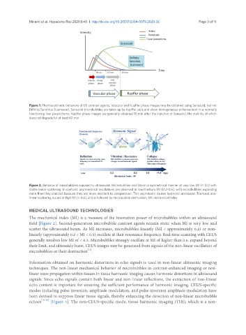
Figure 2. Behavior of microbubbles exposed to ultrasound. Microbubbles oscillate in a symmetrical manner at very low MI (< 0.1) with
stable linear scattering. In contrast, asymmetrical oscillations are observed at low/medium MI (0.2-0.6), with microbubbles expanding
more than they contract because they are more resistant to compression. This asymmetry causes harmonic emissions. Transient non-
linear scattering occurs at high MI (> 0.6), and is followed by microbubble destruction. MI: mechanical index
MEDICAL ULTRASOUND TECHNOLOGIES
The mechanical index (MI) is a measure of the insonation power of microbubbles within an ultrasound
field [Figure 2]. Second-generation microbubble contrast agents remain static when MI is very low and
scatter the ultrasound beam. As MI increases, microbubbles linearly (MI < approximately 0.2) or non-
linearly (approximately 0.2 < MI < 0.5) oscillate at their resonance frequency. Real-time scanning with CEUS
generally involves low MI of < 0.3. Microbubbles strongly oscillate at MI of higher than 0.6, expand beyond
their limit, and ultimately burst. CEUS images may be generated from signals of the non-linear oscillation of
[12]
microbubbles or their destruction .
Information obtained on harmonic distortions in echo signals is used in non-linear ultrasonic imaging
techniques. The non-linear mechanical behavior of microbubbles in contrast-enhanced imaging or non-
linear wave propagation within tissues in tissue harmonic imaging causes harmonic distortions in ultrasound
signals. Since echo signals contain both linear and non-linear reflections, the extraction of non-linear
echo content is important for ensuring the sufficient performance of harmonic imaging. CEUS-specific
modes including pulse inversion, amplitude modulation, and pulse inversion amplitude modulation have
been devised to suppress linear tissue signals, thereby enhancing the detection of non-linear microbubble
echoes [13-15] [Figure 3]. The non-CEUS-specific mode, tissue harmonic imaging (THI), which is a non-