
Page 45 - Read Online
P. 45
Page 6 of 11 Minami et al. Hepatoma Res 2020;6:46 I http://dx.doi.org/10.20517/2394-5079.2020.32
A B C
Figure 7. Defect reperfusion imaging for the local progression of hepatocellular carcinoma (HCC) after radiofrequency ablation (RFA). A:
a computed tomography image shows focal arterial enhancement of the local progression of HCC (arrow) and RFA-induced coagulation
necrosis (arrow heads); B: viable HCC (arrow) in close proximity to the necrotic area (arrow heads) are shown as areas with defects in
the Kupffer phase; C: recurrent HCC (arrow) is clearly identified using the Sonazoid reinjection technique, whereas the necrotic area (arrow
heads) does not enhance
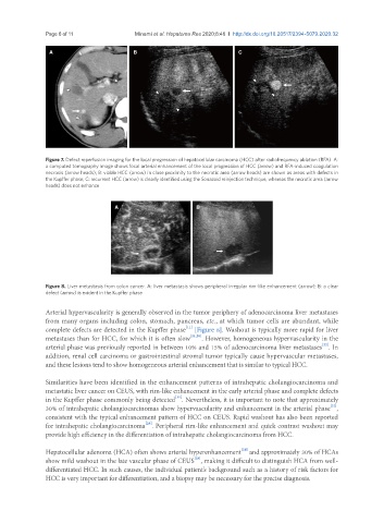
A B
Figure 8. Liver metastasis from colon cancer. A: liver metastasis shows peripheral irregular rim-like enhancement (arrow); B: a clear
defect (arrow) is evident in the Kupffer phase
Arterial hypervascularity is generally observed in the tumor periphery of adenocarcinoma liver metastases
from many organs including colon, stomach, pancreas, etc., at which tumor cells are abundant, while
[11]
complete defects are detected in the Kupffer phase [Figure 8]. Washout is typically more rapid for liver
metastases than for HCC, for which it is often slow [21,28] . However, homogeneous hypervascularity in the
[22]
arterial phase was previously reported in between 10% and 15% of adenocarcinoma liver metastases . In
addition, renal cell carcinoma or gastrointestinal stromal tumor typically cause hypervascular metastases,
and these lesions tend to show homogeneous arterial enhancement that is similar to typical HCC.
Similarities have been identified in the enhancement patterns of intrahepatic cholangiocarcinoma and
metastatic liver cancer on CEUS, with rim-like enhancement in the early arterial phase and complete defects
[11]
in the Kupffer phase commonly being detected . Nevertheless, it is important to note that approximately
[22]
30% of intrahepatic cholangiocarcinomas show hypervascularity and enhancement in the arterial phase ,
consistent with the typical enhancement pattern of HCC on CEUS. Rapid washout has also been reported
[29]
for intrahepatic cholangiocarcinoma . Peripheral rim-like enhancement and quick contrast washout may
provide high efficiency in the differentiation of intrahepatic cholangiocarcinoma from HCC.
[28]
Hepatocellular adenoma (HCA) often shows arterial hyperenhancement and approximately 30% of HCAs
[28]
show mild washout in the late vascular phase of CEUS , making it difficult to distinguish HCA from well-
differentiated HCC. In such causes, the individual patient’s background such as a history of risk factors for
HCC is very important for differentiation, and a biopsy may be necessary for the precise diagnosis.