
Page 46 - Read Online
P. 46
Minami et al. Hepatoma Res 2020;6:46 I http://dx.doi.org/10.20517/2394-5079.2020.32 Page 7 of 11
A B C
Figure 9. Hepatic hemangioma. A: early arterial phase image shows the typical “peripheral globular enhancement” (arrow) of the lesion;
B: the lesion shows “partial centripetal filling” (arrow heads) during the portal phase; C: a progressive filling pattern (arrow heads) is
observed in the Kupffer phase
A B
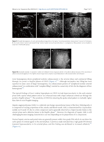
Figure 10. Dysplastic nodule. A: dysplastic nodule (arrowheads) shows hypovascularity in the arterial phase because of the reduction of
arterial and portal supplies; B: the Kupffer phase image shows a slightly hypointense lesion with unclear border (arrowheads)
Liver hemangioma shows peripheral nodular enhancement in the arterial phase and centripetal filling
[11]
through the portal to Kupffer phases of CEUS [Figure 9] . Although incomplete late filling has been
reported in some cases of larger hemangioma, the enhancement pattern of “peripheral nodular arterial
enhancement” in combination with “complete filling” resulted in a sensitivity of 98% for the diagnosis of liver
[30]
hemangioma .
The typical findings of focal nodular hyperplasia on CEUS include hypervascularity in the early arterial
phase with a spoke-wheel pattern and an iso-enhanced mass with a hypo-enhanced central scar through the
[11]
portal to Kupffer phases . The sensitivity of CEUS for detecting the spoke-wheel pattern is markedly higher
than that of color-Doppler imaging.
Hepatic angiomyolipoma (AML) is a relatively rare benign mesenchymal tumor of the liver. Pathologically, it
is composed of varying proportions of fat, muscle, and blood vessels. AML is characterized by a hyperechoic
nodule on B-mode US due to a fatty component and is generally observed as a hypervascular lesion in the
[4]
arterial phase and a defective lesion in the Kupffer phase . However, the diagnosis of hepatic AML is still
challenging because imaging characteristics can vary depending on the proportions of its components.
Normal hepatic arteries and portal veins are generally present within low-grade DNs which do not show the
early uptake of contrast agents in the arterial phase. A previous study indicated that a high-grade DN showed
[11]
transient hypovascularity in the arterial phase, and this finding was attributed to increased cellularity
[Figure 10].