
Page 44 - Read Online
P. 44
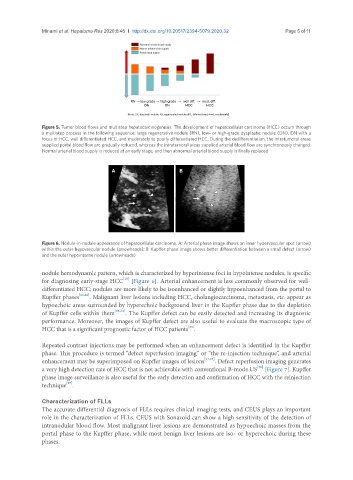
Minami et al. Hepatoma Res 2020;6:46  I  http://dx.doi.org/10.20517/2394-5079.2020.32                                         Page 5 of 11


               Figure 5. Tumor blood flows and multistep hepatocarcinogenesis. The development of hepatocellular carcinoma (HCC) occurs through
               a multistep process in the following sequence: large regenerative nodule (RN), low- or high-grade dysplastic nodule (DN), DN with a
               focus of HCC, well differentiated HCC, and moderately to poorly differentiated HCC. During the dedifferentiation, the intratumoral areas
               supplied portal blood flow are gradually reduced, whereas the intratumoral areas supplied arterial blood flow are synchronously changed.
               Normal arterial blood supply is reduced at an early stage, and then abnormal arterial blood supply is finally replaced


                                       A                      B


               Figure 6. Nodule-in-nodule appearance of hepatocellular carcinoma. A: Arterial phase image shows an inner hypervascular spot (arrow)
               within the outer hypovascular nodule (arrowheads); B: Kupffer phase image shows better differentiation between a small defect (arrow)
               and the outer hypointense nodule (arrowheads)

               nodule hemodynamic pattern, which is characterized by hyperintense foci in hypointense nodules, is specific
                                           [20]
               for diagnosing early-stage HCC  [Figure 6]. Arterial enhancement is less commonly observed for well-
               differentiated HCC; nodules are more likely to be isoenhanced or slightly hypoenhanced from the portal to
               Kupffer phases [21,22] . Malignant liver lesions including HCC, cholangiocarcinoma, metastasis, etc. appear as
               hypoechoic areas surrounded by hyperechoic background liver in the Kupffer phase due to the depletion
               of Kupffer cells within them [11,12] . The Kupffer defect can be easily detected and increasing its diagnostic
               performance. Moreover, the images of Kupffer defect are also useful to evaluate the macroscopic type of
                                                                [23]
               HCC that is a significant prognostic factor of HCC patients .

               Repeated contrast injections may be performed when an enhancement defect is identified in the Kupffer
               phase. This procedure is termed “defect reperfusion imaging” or “the re-injection technique”, and arterial
               enhancement may be superimposed on Kupffer images of lesions [24,25] . Defect reperfusion imaging generates
                                                                                        [26]
               a very high detection rate of HCC that is not achievable with conventional B-mode US  [Figure 7]. Kupffer
               phase image surveillance is also useful for the early detection and confirmation of HCC with the reinjection
                       [27]
               technique .

               Characterization of FLLs
               The accurate differential diagnosis of FLLs requires clinical imaging tests, and CEUS plays an important
               role in the characterization of FLLs. CEUS with Sonazoid can show a high sensitivity of the detection of
               intranodular blood flow. Most malignant liver lesions are demonstrated as hypoechoic masses from the
               portal phase to the Kupffer phase, while most benign liver lesions are iso- or hyperechoic during these
               phases.