
Page 43 - Read Online
P. 43
Page 4 of 11 Minami et al. Hepatoma Res 2020;6:46 I http://dx.doi.org/10.20517/2394-5079.2020.32
A B
Figure 3. Pulse inversion and amplitude modulation. A: pulse-inversion technique is used in second harmonic imaging. Pulse 1 excites
microbubbles, generating a linear fundamental response along with higher harmonic components. The inverted pulse 2 generates the
same frequency components, however with different phases. The linear fundamental response from tissue experiences a 180˚ phase
shift relative to the pulse 1 components, whereas the second harmonic response from microbubble experiences a 360˚ (= 0˚) phase
shift. As a result, the fundamental responses are canceled out and the second harmonic responses are constructively added together;
B: an amplitude modulation technique also plays a role in ultrasonic nonlinear imaging. An amplitude pulse is transmitted to eliminate
the linear response and to elicit a nonlinear response. Upon reception, the pulse 2 components are rescaled and subtracted. Then, the
fundamental response from tissue is canceled, and the second harmonic response from microbubble is leaked out
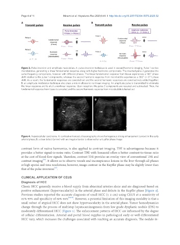
A B
Figure 4. Hepatocellular carcinoma. A: contrast-enhanced ultrasonography shows homogenous strong enhancement (arrow) in the early
arterial phase; B: a clear defect (arrow) with an irregular border is observed on a Kupffer phase image
contrast form of native harmonics, is also applied to contrast imaging. THI is advantageous because it
provides a better signal-to-noise ratio. Contrast THI with Sonazoid offers a better contrast-to-tissue ratio
at the cost of blood flow signals. Therefore, contrast THI provides an overlay view of conventional THI and
[16]
contrast imaging . It allows us to observe vessels and inconspicuous lesions in the liver through all phases
at high spatial and time resolutions; however, image contrast in the Kupffer phase may be slightly lower than
[17]
that of the pulse inversion .
CLINICAL APPLICATION OF CEUS
Diagnosis of HCC
Classic HCC generally receive a blood supply from abnormal arteries alone and are diagnosed based on
positive enhancement (hypervascularity) in the arterial phase and defects in the Kupffer phase [Figure 4].
Previous studies reported the accurate diagnosis of small HCC (≤ 2 cm) using CEUS at a sensitivity of
81%-95% and specificity of 82%-86% [18,19] . However, a potential limitation of this imaging modality is that a
small subset of atypical HCC does not show hypervascularity in the arterial phase. Tumor hemodynamics
change through the process of multistep hepatocarcinogenesis from low grade dysplastic nodule (DN) to
moderately differentiated HCC [Figure 5]. The enhancement patterns of HCC are influenced by the degree
of cellular differentiation. Arterial and portal blood supplies in pathological early or well-differentiated
HCC vary, which increases the challenges associated with reaching an accurate diagnosis. The nodule-in-