
Page 31 - Read Online
P. 31
Page 6 of 14 Kobayashi et al. Hepatoma Res 2020;6:36 I http://dx.doi.org/10.20517/2394-5079.2020.24
A B
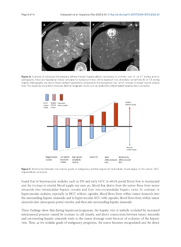
Figure 6. Example of advanced (moderately differentiated) hepatocellular carcinoma in cirrhotic liver. A: on CT during arterial
portography, there are hypodense nodule compared to background liver, which represent loss of nodular portal flow; B: on CT during
hepatic arteriography, the lesion shows marked hyperdensity compared to the background liver, which indicate increased nodular arterial
flow. This hypervascular pattern indicates definite malignant nodule such as moderately differentiated hepatocellular carcinoma
Figure 7. Relationship between the nodular grade of malignancy and the degree of intranodular blood supply of the nodule. HCC:
hepatocellular carcinoma
found that in hypovascular nodules, such as DN and early HCC in which portal blood flow is maintained
and the increase in arterial blood supply not seen yet, blood that drains from the tumor flows from tumor
sinusoids into intranodular hepatic venules and then into extranodular hepatic veins. In contrast, in
hypervascular nodules, especially in HCC without capsules, blood flows from within tumor sinusoids into
the surrounding hepatic sinusoids; and in hypervascular HCC with capsules, blood flows from within tumor
sinusoids into intracapsule portal venules, and then into surrounding hepatic sinusoids.
These findings show that during hepatocarcinogenesis, the hepatic vein is initially occluded by increased
intratumoral pressure caused by increase in cell density, and direct connections between tumor sinusoids
and surrounding hepatic sinusoids work as the tumor drainage route because of occlusion of the hepatic
vein. Then, as the nodular grade of malignancy progresses, the tumor becomes encapsulated and the direct