
Page 29 - Read Online
P. 29
Page 4 of 14 Kobayashi et al. Hepatoma Res 2020;6:36 I http://dx.doi.org/10.20517/2394-5079.2020.24
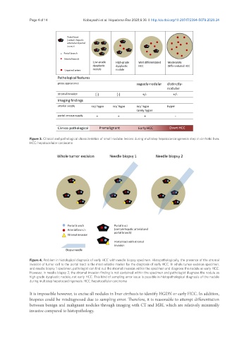
Figure 3. Clinical and pathological characteristics of small nodular lesions during multistep hepatocarcinogenesis step in cirrhotic liver.
HCC: hepatocellular carcinoma
Figure 4. Problem in histological diagnosis of early HCC with needle biopsy specimen. Histopathologically, the presence of the stromal
invasion of tumor cell to the portal tract is the most reliable marker for the diagnosis of early HCC. In whole tumor excision specimen,
and needle biopsy 1 specimen, pathologist can find out the stromal invasion within the specimen and diagnose the nodule as early HCC.
However, in needle biopsy 2, the stromal invasion finding is not contained within the specimen and pathologist diagnose the nodule as
high-grade dysplastic nodule, not early HCC. This kind of sampling error issue is possible in histopathological diagnosis of the nodule
during multistep hepatocarcinigenesis. HCC: hepatocellular carcinoma
It is impossible however, to excise all nodules in liver cirrhosis to identify HGDN or early HCC. In addition,
biopsies could be misdiagnosed due to sampling error. Therefore, it is reasonable to attempt differentiation
between benign and malignant nodules through imaging with CT and MRI, which are relatively minimally
invasive compared to histopathology.