
Page 176 - Read Online
P. 176
Page 8 of 15 Rong et al. Vessel Plus 2019;3:18 I http://dx.doi.org/10.20517/2574-1209.2019.007
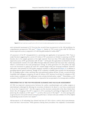
Figure 5. 3D semi-automatic quantification of the left ventricle demonstrating global function and regional wall motion
semi-automated assessment of LV function has recently been incorporated in the ASE guidelines for
[31]
comprehensive perioperative TEE exam [Figure 5]. Similar to TTE, recent studies with 3D TEE have
[32]
shown improved accuracy compared to 2D with the gold standard of cardiac MRI .
3D assessment of the RV intraoperatively is a promising new application of perioperative TEE. There is
ample literature suggesting that current 2D indices of RV assessment are limited in assessing overall RV
function due to the complex geometry of the right ventricle. There have been TTE studies demonstrating
[19]
improved accuracy of RV quantification with 3D echo . In the past, this application was limited by the fact
that measurements needed to be made offline through dedicated software and were more time consuming,
which is cumbersome in the acute perioperative setting. In earlier generation 3D TEE probes, the large
sector size necessary to capture the entire RV created images with poor temporal resolution. However,
with newer, more powerful 3D TEE probes, temporal resolution is often preserved [Figure 6] in addition to
providing superior spatial resolution. Though 3D RV evaluation is feasible, there remains few intraoperative
RV 3D imaging studies to date, and those that exist performed analyses off-line. Of note, a recent article by
Grønlykke and colleagues comparing 2D and 3D indices of RV function found that 3D evaluation of RV
stroke volume correlated with RV pulmonary artery catheter derived cardiac output . Nevertheless, as 3D
[33]
evaluation of the RV becomes more efficient, it is expected to be incorporated to the intraoperative workflow.
PERIOPERATIVE 3D TEE FOR PROCEDURE GUIDANCE AND VALVULAR ASSESSMENT
3D TEE has improved communication between the cardiac anesthesiologist and cardiac surgeon or
interventional cardiologist by allowing the structures of interest to be shown in real-time, reconstructed
in the en face “surgeon’s view” - how the surgeon sees the structure anatomically in the chest. Studies have
shown that 3D imaging of the MV is superior to 2D in assessing the pathology of degenerative MR and
[37]
predicting the complexity of MV repair [34-36] , as well as the success of the repair . As valve sparing aortic
surgeries become more preferred and commonly performed, it remains to be seen whether 3D will be helpful
in predicting success of aortic valve repairs .
[38]
Advancements in 3D technology has allowed real time 3D TEE to have a central role in percutaneous
structural heart interventions. While guidance during these procedures was originally recommended