
Page 172 - Read Online
P. 172
Page 4 of 15 Rong et al. Vessel Plus 2019;3:18 I http://dx.doi.org/10.20517/2574-1209.2019.007
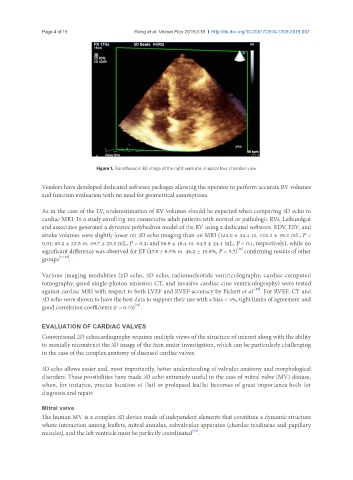
Figure 1. Transthoracic 3D image of the right ventricle in apical four chamber view
Vendors have developed dedicated software packages allowing the operator to perform accurate RV volumes
and function evaluation with no need for geometrical assumptions.
As in the case of the LV, underestimation of RV volumes should be expected when comparing 3D echo to
cardiac MRI. In a study enrolling 100 consecutive adult patients with normal or pathologic RVs, Leibundgut
and associates generated a dynamic polyhedron model of the RV using a dedicated software. EDV, ESV, and
stroke volumes were slightly lower on 3D echo imaging than on MRI (124.0 ± 34.4 vs. 134.2 ± 39.2 mL, P <
0.01; 65.2 ± 23.5 vs. 69.7 ± 25.5 mL, P = 0.2; and 58.8 ± 18.4 vs. 64.5 ± 24.1 mL, P < 0.1, respectively), while no
[16]
significant difference was observed for EF (47.8 ± 8.5% vs. 48.2 ± 10.8%, P = 5.7) confirming results of other
groups [17,18] .
Various imaging modalities (2D echo, 3D echo, radionucleotide ventriculography, cardiac computed
tomography, gated single-photon emission CT, and invasive cardiac cine ventriculography) were tested
[19]
against cardiac MRI with respect to both LVEF and RVEF accuracy by Pickett et al. . For RVEF, CT and
3D echo were shown to have the best data to support their use with a bias < 5%, tight limits of agreement and
[19]
good correlation coefficients (r > 0.75) .
EVALUATION OF CARDIAC VALVES
Conventional 2D echocardiography requires multiple views of the structure of interest along with the ability
to mentally reconstruct the 3D image of the item under investigation, which can be particularly challenging
in the case of the complex anatomy of diseased cardiac valves.
3D echo allows easier and, most importantly, better understanding of valvular anatomy and morphological
disorders. These possibilities have made 3D echo extremely useful in the case of mitral valve (MV) disease,
when, for instance, precise location of flail or prolapsed leaflet becomes of great importance both for
diagnosis and repair.
Mitral valve
The human MV is a complex 3D device made of independent elements that constitute a dynamic structure
where interaction among leaflets, mitral annulus, subvalvular apparatus (chordae tendineae and papillary
[20]
muscles), and the left ventricle must be perfectly coordinated .