
Page 167 - Read Online
P. 167
Page 2 of 3 Ryabova et al. Vessel Plus 2019;3:17 I http://dx.doi.org/10.20517/2574-1209.2019.10
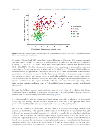
Figure 1. Distribution of protoporfyrine IX (PpIX) and MitoTracker fluorescence intensity in cybrid lines and THP-1 macrophage lines after
4 h of incubation with 5-aminolevulinic acid
We studied 5-ALA-induced PpIX accumulation on cybrid lines derived from rho0 THP-1 macrophages and
patients’ thrombocytes with mutation-bearing proatherosclerotic mitochondria. We used 3 cybrid lines (TC-
HSMAM1, TC-HSM1, TC-LSM1) and 2 native THP-1 monocyte cultures obtained from different sources
(THP-1 IBG, THP-1 GPI). The mitochondrial functional state was assessed as mitochondrial membrane
TM
potential using fluorescence intensity of mitochondrial dye MitoTracker Orange CMTMRos (Thermo
Fisher Scientific). A high level of fluorescence is characteristic of active mitochondria; on the contrary,
dysfunctional mitochondria possess a low level of fluorescence. Estimation of fluorescence was performed by
laser scanning microscopy with spectral unmixing. MitoTracker and PpIX dyes were excited with a 561 nm
laser, and fluorescence was detected at 570-750 nm wavelength. The signal was spectrally unmixed to obtain
separate MitoTracker and PpIX signals. Individual cells areas were traced on microscopic images taken
under identical conditions, and mean values of PpIX and MitoTracker fluorescence intensity were obtained
for each cell.
We found that PpIX accumulation varied significantly from cell to cell within cell populations. Cybrid lines
had increased PpIX accumulation in comparison with native THP-1 macrophage lines. A positive correlation
between PpIX and mitochondrial potential was observed [Figure 1].
It can be assumed that selective elimination of dysfunctional (mutated) mitochondria can be achieved
by adjusting laser intensity sufficient to induce photodynamic destruction of the organelles, while active
(normal) mitochondria and the cells as a whole should be preserved at the used intensities.
Photodynamic therapy is a non-invasive treatment method in oncology, dermatology and infectious
diseases. The disadvantage of this approach is the low depth of penetration into the tissue of excitation light
and insufficient accumulation of photosensitizers in subcellular organelles. On the other hand, subcellular
organelles, and in particular mitochondria, are promising therapeutic targets for effective therapy. At the
same time, targeting mitochondria approaches do not necessarily have to be associated with the use of
laser. It can be assumed that substances accumulating in mitochondria as a result of metabolic disturbances
themselves without a laser will contribute to the death of dysfunctional mitochondria if there is an intra-
mitochondrial overload of such substances. In addition to 5-ALA, there are many other mitochondria