
Page 175 - Read Online
P. 175
Rong et al. Vessel Plus 2019;3:18 I http://dx.doi.org/10.20517/2574-1209.2019.007 Page 7 of 15
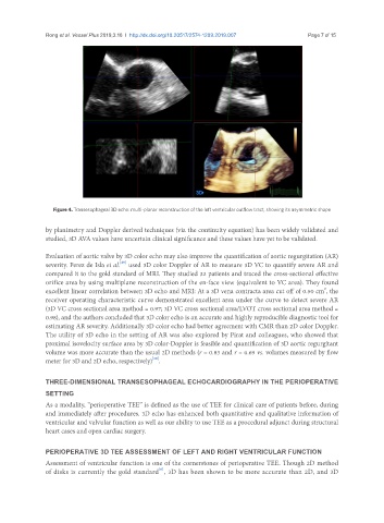
Figure 4. Transesophageal 3D echo: multi-planar reconstruction of the left ventricular outflow tract, showing its asymmetric shape
by planimetry and Doppler derived techniques (via the continuity equation) has been widely validated and
studied, 3D AVA values have uncertain clinical significance and these values have yet to be validated.
Evaluation of aortic valve by 3D color echo may also improve the quantification of aortic regurgitation (AR)
[29]
severity. Perez de Isla et al. used 3D color Doppler of AR to measure 3D VC to quantify severe AR and
compared it to the gold standard of MRI. They studied 32 patients and traced the cross-sectional effective
orifice area by using multiplane reconstruction of the en-face view (equivalent to VC area). They found
2
excellent linear correlation between 3D echo and MRI: At a 3D vena contracta area cut off of 0.50 cm , the
receiver operating characteristic curve demonstrated excellent area under the curve to detect severe AR
(3D VC cross sectional area method = 0.97; 3D VC cross sectional area/LVOT cross sectional area method =
0.98), and the authors concluded that 3D color echo is an accurate and highly reproducible diagnostic tool for
estimating AR severity. Additionally 3D color echo had better agreement with CMR than 2D color Doppler.
The utility of 3D echo in the setting of AR was also explored by Pirat and colleagues, who showed that
proximal isovelocity surface area by 3D color-Doppler is feasible and quantification of 3D aortic regurgitant
volume was more accurate than the usual 2D methods (r = 0.83 and r = 0.69 vs. volumes measured by flow
[30]
meter for 3D and 2D echo, respectively) .
THREE-DIMENSIONAL TRANSESOPHAGEAL ECHOCARDIOGRAPHY IN THE PERIOPERATIVE
SETTING
As a modality, “perioperative TEE” is defined as the use of TEE for clinical care of patients before, during
and immediately after procedures. 3D echo has enhanced both quantitative and qualitative information of
ventricular and valvular function as well as our ability to use TEE as a procedural adjunct during structural
heart cases and open cardiac surgery.
PERIOPERATIVE 3D TEE ASSESSMENT OF LEFT AND RIGHT VENTRICULAR FUNCTION
Assessment of ventricular function is one of the cornerstones of perioperative TEE. Though 2D method
[6]
of disks is currently the gold standard , 3D has been shown to be more accurate than 2D, and 3D