
Page 179 - Read Online
P. 179
Rong et al. Vessel Plus 2019;3:18 I http://dx.doi.org/10.20517/2574-1209.2019.007 Page 11 of 15
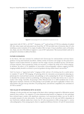
Figure 9. Transesophageal 3D-color assessment of a paravalvular leak
[52]
[53]
respectively) with 3D TEE vs. 2D TEE . Kronzon et al. used real time 3D TEE for evaluation of residual
MR after mitral repair and replacement and found that 3D TEE provided more information than 2D echo
including accurate evaluation of the origin of residual MR, the type of ring or prosthesis used, and location,
size, shape, and area of the dehisced segment in cases of MV replacement. This increased detail could aid in
perioperative decisions to address the defect.
FUTURE SCENARIOS
In the future, TEE may routinely be combined with fluoroscopy for multimodality real-time fusion
guidance during interventional procedures. Indeed, overlay of markers and images has the potential to
improve communication between the operator and the imager, increase procedural success, and decrease
radiation exposure. Future prospective studies are necessary to evaluate the utility of live fusion imaging.
In addition, the advent of 3D capabilities of intracardiac echocardiographic catheters suggests that this
[54]
imaging modality should be revisited for potential guidance during catheter-based interventions .
Until live fusion or “augmented reality” imaging is fully realized, 3D printing may be a useful option
to translate CT and 3D TEE imaging. 3D printing allows for discussion and preoperative planning on
patient-specific structural heart anatomy and pathology as well as preparing for 2D and 3D imaging best
to elucidate the anatomy and device positioning. 3D printing may also help select device size in cases of
unclear valve sizing. Currently, 3D printing is often obtained from CT or MRI modalities due to their
high spatial resolution. However, 3D TEE printing is now possible, and may be applicable to future valve
interventions after fine tuning work flow, 3D printing times, and software compatibility for conversion of
3D TEE to models .
[55]
THE VALUE OF EXPERIENCE WITH 3D ECHO
Although 3D echo provides real-time images of the heart valves, training is required to differentiate normal
anatomy from artifacts. The majority of studies demonstrating benefits in diagnosis of valve pathology
have included expert readers with significant experience. Less experienced readers may not be able to
accurately interpret 3D echo information. The utility and accuracy of 3D echo and MPR is dependent on
user familiarity; multiple studies have shown that 3D echo measurements differ among expert vs. novice
echocardiographers. Even among experts, there is a significant difference between 2D and 3D measurements