
Page 178 - Read Online
P. 178
Page 10 of 15 Rong et al. Vessel Plus 2019;3:18 I http://dx.doi.org/10.20517/2574-1209.2019.007
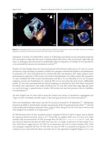
Figure 8. Transesophageal 3D view of the double orifice created by MitraClip, a percutaneous mitral valve repair device (A); quantitative
assessment of valve area of the double orifice by MPR; Multi-planar reconstruction (B)
Assessment of severity of residual MR is central as it determines procedural success and patient prognosis
after percutaneous edge-edge clip repair. Grading residual MR severity after percutaneous edge-edge clip
repair is challenging and assessment has traditionally relied on integration of multiple 2D echo parameters
as recommended by national and international societies [43,44] .
Notably, 3D color Doppler allows for direct measurement of the effective orifice area or VC area. In a recent
retrospective study enrolling 155 patients, Avenatti and colleagues evaluated the feasibility and performance
of summative VC area of multiple jets for residual MR after percutaneous MV repair against expert
multiparametric appraisal of MR severity and invasive hemodynamics; the authors found that summative
2
VC area correlated well with invasive hemodynamics and that a VC area threshold of 0.27 cm had good
diagnostic accuracy for identification of ≥ moderate MR with an area under the curve of 0.81. Additionally,
smaller VC area were associated with less clinical symptoms as measured by New York Heart Association
functional class improvement. This study introduces total VC area after mitral valve edge-edge clip repair
as a novel technique in quantification of residual MR severity and may have potential value in Guidelines
[45]
recommendations .
3D color Doppler has also been used to assess the location and amount of paravalvular regurgitation jets
[46]
[Figure 9] both immediately and after repair, or later during paravalvular leak closure procedures .
[42]
Valve area immediately after repair can also be accurately assessed by 3D planimetry . Multiplanar
reformatting (MPR) is used for linear annular measurements of the tricuspid and mitral valves [47,48] and aids
in the periprocedural sizing of rings and valves, sizing of ASDs, sizing of LAA for LAA closure devices, and
valve sizing for transcatheter valve replacement [28,49,50] .
3D TEE has been shown to have increased accuracy compared to 2D echo which in many cases is relevant
[51]
for improved clinical outcomes. Johri et al. found that in complex ASDs (33% of a total of 24 ASDs
studied), ASD area measured by 3D TEE was larger than 2D TEE (2.8 ± 1.3 vs. 1.7 ± 1.4 cm ; P < 0.05). This
2
was clinically important because 3D TEE areas were also 27% larger in patients that had residual shunt
after ASD closure suggesting that 2D TEE can underestimate the area of complex-shaped ASDs. Streb
and colleagues compared real time 2D TEE with real time 3D TEE in 40 patients during place of a LAA
occlusion device and found that there was better device size agreement (weighted Kappa 0.62 vs. 0.28,