
Page 173 - Read Online
P. 173
Rong et al. Vessel Plus 2019;3:18 I http://dx.doi.org/10.20517/2574-1209.2019.007 Page 5 of 15
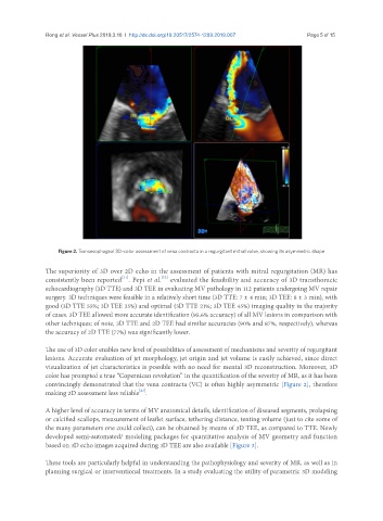
Figure 2. Transesophageal 3D-color assessment of vena contracta in a regurgitant mitral valve, showing its asymmetric shape
The superiority of 3D over 2D echo in the assessment of patients with mitral regurgitation (MR) has
[22]
[21]
consistently been reported . Pepi et al. evaluated the feasibility and accuracy of 3D transthoracic
echocardiography (3D TTE) and 3D TEE in evaluating MV pathology in 112 patients undergoing MV repair
surgery. 3D techniques were feasible in a relatively short time (3D TTE: 7 ± 4 min; 3D TEE: 8 ± 3 min), with
good (3D TTE 55%; 3D TEE 35%) and optimal (3D TTE 21%; 3D TEE 45%) imaging quality in the majority
of cases. 3D TEE allowed more accurate identification (95.6% accuracy) of all MV lesions in comparison with
other techniques; of note, 3D TTE and 2D TEE had similar accuracies (90% and 87%, respectively), whereas
the accuracy of 2D TTE (77%) was significantly lower.
The use of 3D color enables new level of possibilities of assessment of mechanisms and severity of regurgitant
lesions. Accurate evaluation of jet morphology, jet origin and jet volume is easily achieved, since direct
visualization of jet characteristics is possible with no need for mental 3D reconstruction. Moreover, 3D
color has prompted a true “Copernican revolution” in the quantification of the severity of MR, as it has been
convincingly demonstrated that the vena contracta (VC) is often highly asymmetric [Figure 2], therefore
[23]
making 2D assessment less reliable .
A higher level of accuracy in terms of MV anatomical details, identification of diseased segments, prolapsing
or calcified scallops, measurement of leaflet surface, tethering distance, tenting volume (just to cite some of
the many parameters one could collect), can be obtained by means of 3D TEE, as compared to TTE. Newly
developed semi-automated/ modeling packages for quantitative analysis of MV geometry and function
based on 3D echo images acquired during 3D TEE are also available [Figure 3].
These tools are particularly helpful in understanding the pathophysiology and severity of MR, as well as in
planning surgical or interventional treatments. In a study evaluating the utility of parametric 3D modeling