


Page 66 - Read Online
P. 66
Page 10 of 19                 Rinaldi et al. Vessel Plus 2024;8:21  https://dx.doi.org/10.20517/2574-1209.2023.65


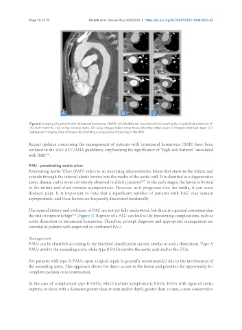
                Figure 6. Imaging of a patient with intramural hematoma (IMH). (A) Multiplanar reconstruction revealing the longitudinal extension of
                the IMH from the LSA to the visceral aorta. (B) Axial images taken a few hours after the initial onset of thoracic and back pain. (C)
                Subsequent imaging after 48 hours documenting a progressive thickening of the IMH.

               Recent updates concerning the management of patients with intramural hematoma (IMH) have been
               outlined in the 2022 ACC/AHA guidelines, emphasizing the significance of “high-risk features” associated
                        [46]
               with IMH .
               PAU - penetrating aortic ulcer
               Penetrating Aortic Ulcer (PAU) refers to an ulcerating atherosclerotic lesion that starts in the intima and
               extends through the internal elastic lamina into the media of the aortic wall. It is classified as a degenerative
               aortic disease and is more commonly observed in elderly patients . In the early stages, the lesion is limited
                                                                      [47]
               to the intima and often remains asymptomatic. However, as it progresses into the media, it can cause
               thoracic pain. It is important to note that a significant number of patients with PAU may remain
               asymptomatic, and these lesions are frequently discovered incidentally.


               The natural history and evolution of PAU are not yet fully understood, but there is a general consensus that
                                     [48]
               the risk of rupture is high  [Figure 7]. Rupture of a PAU can lead to life-threatening complications, such as
               aortic dissection or intramural hematoma. Therefore, prompt diagnosis and appropriate management are
               essential in patients with suspected or confirmed PAU.

               Management
               PAUs can be classified according to the Stanford classification system, similar to aortic dissections. Type A
               PAUs involve the ascending aorta, while type B PAUs involve the aortic arch and/or the DTA.

               For patients with type A PAUs, open surgical repair is generally recommended due to the involvement of
               the ascending aorta. This approach allows for direct access to the lesion and provides the opportunity for
               complete excision or reconstruction.


               In the case of complicated type B PAUs, which include symptomatic PAUs, PAUs with signs of aortic
               rupture, or those with a diameter greater than 20 mm and/or depth greater than 10 mm, a non-conservative

