


Page 62 - Read Online
P. 62
Page 6 of 19                  Rinaldi et al. Vessel Plus 2024;8:21  https://dx.doi.org/10.20517/2574-1209.2023.65


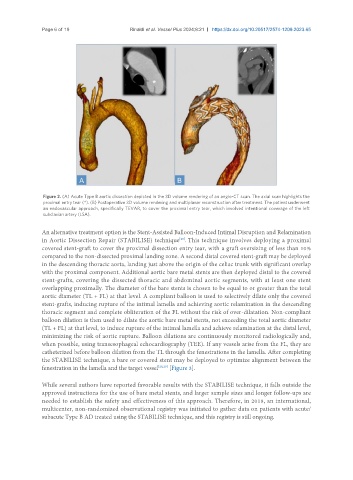
                Figure 2. (A) Acute Type B aortic dissection depicted in the 3D volume rendering of an angio-CT scan. The axial scan highlights the
                proximal entry tear (*). (B) Postoperative 3D volume rendering and multiplanar reconstruction after treatment. The patient underwent
                an endovascular approach, specifically TEVAR, to cover the proximal entry tear, which involved intentional coverage of the left
                subclavian artery (LSA).

               An alternative treatment option is the Stent-Assisted Balloon-Induced Intimal Disruption and Relamination
                                                              [28]
               in Aortic Dissection Repair (STABILISE) technique . This technique involves deploying a proximal
               covered stent-graft to cover the proximal dissection entry tear, with a graft oversizing of less than 10%
               compared to the non-dissected proximal landing zone. A second distal covered stent-graft may be deployed
               in the descending thoracic aorta, landing just above the origin of the celiac trunk with significant overlap
               with the proximal component. Additional aortic bare metal stents are then deployed distal to the covered
               stent-grafts, covering the dissected thoracic and abdominal aortic segments, with at least one stent
               overlapping proximally. The diameter of the bare stents is chosen to be equal to or greater than the total
               aortic diameter (TL + FL) at that level. A compliant balloon is used to selectively dilate only the covered
               stent-grafts, inducing rupture of the intimal lamella and achieving aortic relamination in the descending
               thoracic segment and complete obliteration of the FL without the risk of over-dilatation. Non-compliant
               balloon dilation is then used to dilate the aortic bare metal stents, not exceeding the total aortic diameter
               (TL + FL) at that level, to induce rupture of the intimal lamella and achieve relamination at the distal level,
               minimizing the risk of aortic rupture. Balloon dilations are continuously monitored radiologically and,
               when possible, using transesophageal echocardiography (TEE). If any vessels arise from the FL, they are
               catheterized before balloon dilation from the TL through the fenestrations in the lamella. After completing
               the STABILISE technique, a bare or covered stent may be deployed to optimize alignment between the
               fenestration in the lamella and the target vessel [28,29]  [Figure 3].

               While several authors have reported favorable results with the STABILISE technique, it falls outside the
               approved instructions for the use of bare metal stents, and larger sample sizes and longer follow-ups are
               needed to establish the safety and effectiveness of this approach. Therefore, in 2018, an international,
               multicenter, non-randomized observational registry was initiated to gather data on patients with acute/
               subacute Type B AD treated using the STABILISE technique, and this registry is still ongoing.

