


Page 61 - Read Online
P. 61
Rinaldi et al. Vessel Plus 2024;8:21  https://dx.doi.org/10.20517/2574-1209.2023.65   Page 5 of 19


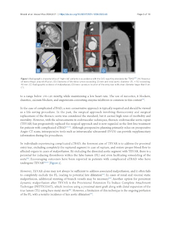
                Figure 1. Radiographic characteristics of “high-risk” patients in accordance with the SVS reporting standards for TBAD [16] . (A) Presence
                of hemorrhagic pleural effusion. (B) Diameter of the false lumen exceeding 22 mm and total aortic diameter (TL + FL) exceeding
                40 mm. (C) Radiographic evidence of malperfusion. (D) Inner curvature location of the entry tear with a tear diameter larger than 1 cm
                (*).

               to a range below 100-120 mmHg while maintaining a low heart rate. The use of narcotics, ß blockers,
               diuretics, calcium blockers, and angiotensin-converting enzyme inhibitors is common in this context .
                                                                                                    [18]

               In the case of complicated aTBAD, a non-conservative approach is typically required and should be viewed
               as a life-saving procedure. In the past, the surgical approach involving thoracotomy and surgical
               replacement of the thoracic aorta was considered the standard, but it carried high rates of morbidity and
               mortality. However, with the advancements in endovascular techniques, thoracic endovascular aortic repair
               (TEVAR) has progressively replaced the surgical approach and is now regarded as the first-line treatment
               for patients with complicated aTBAD [19,20] . Although preoperative planning primarily relies on preoperative
               Angio-CT scans, intraoperative tools such as intravascular ultrasound (IVUS) can provide supplementary
               information during the procedures.

               In individuals experiencing complicated aTBAD, the foremost aim of TEVAR is to address the proximal
               entry tear, excluding completely the ruptured segment in case of rupture, and restore proper blood flow to
               affected organs in cases of malperfusion. By excluding the dissected aortic segment with TEVAR, there is a
               potential for inducing thrombosis within the false lumen (FL) and even facilitating remodeling of the
               aorta . Encouraging outcomes have been reported in patients with complicated aTBAD who have
                   [21]
               undergone TEVAR [22,23]  [Figure 2].

               However, TEVAR alone may not always be sufficient to address associated malperfusion, and it often fails
               to completely exclude the FL, leading to potential late dilatation . In cases of renal and visceral static
                                                                        [24]
                                                                             [25]
               malperfusion, additional stenting of branch vessels may be necessary . Another option for persistent
               dynamic malperfusion after TEVAR is the Provisional Extension To Induce Complete Attachment
               Technique (PETTICOAT), which involves using a proximal stent graft along with distal expansion of the
               true lumen (TL) using bare metal stents . However, a limitation of this technique is the ongoing perfusion
                                                 [26]
               of the FL, with a notable incidence of late aortic dilatation .
                                                                [27]

