


Page 12 - Read Online
P. 12
Hong et al. Vessel Plus 2022;6:4  https://dx.doi.org/10.20517/2574-1209.2021.88  Page 7 of 17


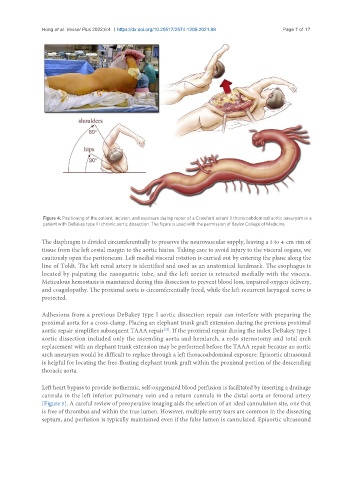
                Figure 4. Positioning of the patient, incision, and exposure during repair of a Crawford extent II thoracoabdominal aortic aneurysm in a
                patient with DeBakey type III chronic aortic dissection. The figure is used with the permission of Baylor College of Medicine.

               The diaphragm is divided circumferentially to preserve the neurovascular supply, leaving a 3 to 4-cm rim of
               tissue from the left costal margin to the aortic hiatus. Taking care to avoid injury to the visceral organs, we
               cautiously open the peritoneum. Left medial visceral rotation is carried out by entering the plane along the
               line of Toldt. The left renal artery is identified and used as an anatomical landmark. The esophagus is
               located by palpating the nasogastric tube, and the left ureter is retracted medially with the viscera.
               Meticulous hemostasis is maintained during this dissection to prevent blood loss, impaired oxygen delivery,
               and coagulopathy. The proximal aorta is circumferentially freed, while the left recurrent laryngeal nerve is
               protected.

               Adhesions from a previous DeBakey type I aortic dissection repair can interfere with preparing the
               proximal aorta for a cross-clamp. Placing an elephant trunk graft extension during the previous proximal
               aortic repair simplifies subsequent TAAA repair . If the proximal repair during the index DeBakey type I
                                                        [32]
               aortic dissection included only the ascending aorta and hemiarch, a redo sternotomy and total arch
               replacement with an elephant trunk extension may be performed before the TAAA repair because an aortic
               arch aneurysm would be difficult to replace through a left thoracoabdominal exposure. Epiaortic ultrasound
               is helpful for locating the free-floating elephant trunk graft within the proximal portion of the descending
               thoracic aorta.


               Left heart bypass to provide isothermic, self-oxygenated blood perfusion is facilitated by inserting a drainage
               cannula in the left inferior pulmonary vein and a return cannula in the distal aorta or femoral artery
               [Figure 5]. A careful review of preoperative imaging aids the selection of an ideal cannulation site, one that
               is free of thrombus and within the true lumen. However, multiple entry tears are common in the dissecting
               septum, and perfusion is typically maintained even if the false lumen is cannulated. Epiaortic ultrasound

