
Page 211 - Read Online
P. 211
Page 12 of 23 Farber et al. Plast Aesthet Res 2020;7:20 I http://dx.doi.org/10.20517/2347-9264.2020.05
Figure 17. Brief algorithm followed for adults with acquired ptosis and good levator function
A B
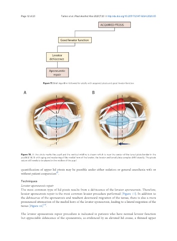
Figure 18. A: the circle marks the pupil and the vertical midline is drawn which is near the center of the tarsal plate border in the
youthful lid; B: with aging and weakening of the medial horn of the levator, the levator and tarsal plate complex shift laterally. The ptosis
suture still needs to be placed in the midline of the pupil
quantification of upper lid ptosis may be possible under either sedation or general anesthesia with or
[9]
without patient cooperation .
Techniques
Levator aponeurosis repair
The most common type of lid ptosis results from a dehiscence of the levator aponeurosis. Therefore,
levator aponeurosis repair is the most common levator procedure performed [Figure 17]. In addition to
the dehiscence of the aponeurosis and resultant downward migration of the tarsus, there is also a more
pronounced attenuation of the medial horn of the levator aponeurosis, leading to a lateral migration of the
[10]
tarsus [Figure 18] .
The levator aponeurosis repair procedure is indicated in patients who have normal levator function
but appreciable dehiscence of the aponeurosis, as evidenced by an elevated lid crease, a thinned upper