Page 215 - Read Online
P. 215
Page 16 of 23                                        Farber et al. Plast Aesthet Res 2020;7:20  I  http://dx.doi.org/10.20517/2347-9264.2020.05


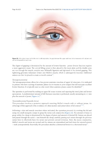
               Figure 23. After ptosis repair while the knot is still adjustable, the gap between the upper and lower lid is measured with calipers and
               can be compared from each side


               The degree of gapping is determined by the amount of levator function - poorer levator function requires
               a more aggressive repair. The central lifting suture is then placed in the tarsal plate and the double arms
               are run through the muscle complex near Whitnall’s ligament and tightened to the desired gapping. This
               tightening generates redundant levator and Müller’s muscle, which is subsequently resected. Additional
                                                             [1]
               sutures can then be placed to create a smooth contour .
               Tarsoaponeurectomy
               The tarsoaponeurectomy allows for a less precise anatomic resection of upper lid structures. It is indicated
               in patients who have scarring of anatomic planes due to trauma or prior surgery but still maintain excellent
                                                                                              [11]
               levator function. It is typically used as a last resort when anatomic planes cannot be identified .
               The operation is performed by making an upper lid crease incision and exposing the tarsal plate and levator
               aponeurosis. A predetermined amount of full thickness resection is performed, usually amounting to 0-2 mm
                                              [1]
               plus the amount of ptosis [Figure 24] .
               Tarsomullerectomy/Fasanella-Servat
               This procedure involves a posterior approach resecting Müller’s muscle with or without tarsus. An
               advantage of this approach is the avoidance of a skin incision and preservation of lid contour [12,13] .

               Following a skin and muscle resection where indicated, the operation proceeds by everting the lid and
               using two small mosquito clamps to grasp the tarsus and conjunctiva [Figure 25]. The amount of tissue to
               grasp within the clamp is determined by the degree of ptosis and amount of desired lift. Sutures are placed
               and passed through the eyelid 1 mm beneath the clamp carefully passing each suture through the previous
               suture hole to effectively bury the suture and prevent corneal irritation. Next, the excess conjunctiva,
               Müller’s muscle and tarsus are excised and the sutures are externalized and tied down for removal around
                                                                                                    [1]
               1 week postoperatively. Essentially, this procedure attaches a disinserted levator to a shortened tarsus .

