
Page 216 - Read Online
P. 216
Farber et al. Plast Aesthet Res 2020;7:20 I http://dx.doi.org/10.20517/2347-9264.2020.05 Page 17 of 23
A
B
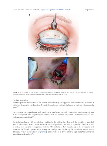
Figure 24. A: markings on the levator aponeurosis and superior tarsal plate for excision of full thickness tissue during a
tarsoaponeurectomy; B: the black eye protector can be seen during a tarsoaponeurectomy
Frontalis suspension
Frontalis procedures circumvent the levator when elevating the upper lid and are therefore indicated in
patients with poor levator function. Typically, frontalis suspension is indicated in patients with congenital
[1]
ptosis .
The operation can be performed with prosthetic or autologous material. Fascia lata is most commonly used
in the adult patient with acquired ptosis. Silicone rods are reserved for pediatric patients who do not have
sufficient donor site fascia .
[1]
The technique begins with a single brow incision in the midpupillary line until the frontalis is identified.
Next, a lid crease incision is made, and the superior edge of the tarsal plate is exposed to allow for sutures
to be tied over a strand of suspensory material. This material is then passed deep to the orbicularis until
it reaches the frontalis, generating a pentagonal configuration by placing the lateral and medial sutures
inferiorly outside of the limbus [Figure 26]. The lid crease is closed prior to tightening the suspensory
[1]
material at the brow level .