
Page 214 - Read Online
P. 214
Farber et al. Plast Aesthet Res 2020;7:20 I http://dx.doi.org/10.20517/2347-9264.2020.05 Page 15 of 23
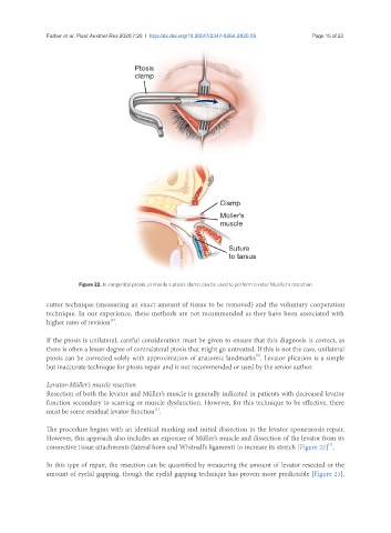
Figure 22. In congenital ptosis, primarily a ptosis clamp can be used to perform levator Mueller’s resection
cutter technique (measuring an exact amount of tissue to be removed) and the voluntary cooperation
technique. In our experience, these methods are not recommended as they have been associated with
[9]
higher rates of revision .
If the ptosis is unilateral, careful consideration must be given to ensure that this diagnosis is correct, as
there is often a lesser degree of contralateral ptosis that might go untreated. If this is not the case, unilateral
[9]
ptosis can be corrected solely with approximation of anatomic landmarks . Levator plication is a simple
but inaccurate technique for ptosis repair and is not recommended or used by the senior author.
Levator-Müller’s muscle resection
Resection of both the levator and Müller’s muscle is generally indicated in patients with decreased levator
function secondary to scarring or muscle dysfunction. However, for this technique to be effective, there
[1]
must be some residual levator function .
The procedure begins with an identical marking and initial dissection to the levator aponeurosis repair.
However, this approach also includes an exposure of Müller’s muscle and dissection of the levator from its
[1]
connective tissue attachments (lateral horn and Whitnall’s ligament) to increase its stretch [Figure 22] .
In this type of repair, the resection can be quantified by measuring the amount of levator resected or the
amount of eyelid gapping, though the eyelid gapping technique has proven more predictable [Figure 23].