
Page 206 - Read Online
P. 206
Farber et al. Plast Aesthet Res 2020;7:20 I http://dx.doi.org/10.20517/2347-9264.2020.05 Page 7 of 23
A B
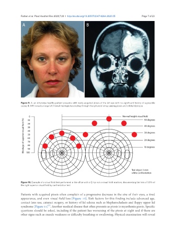
Figure 9. A: an otherwise healthy patient presents with newly acquired ptosis of the left eye with no significant history of a possible
cause; B: MRI reveals a large left frontal meningioma eroding through the sphenoid wing causing ptosis and orbital dystopia
Figure 10. Example of a visual field test performed in the office with a Q-tip not a visual field machine, documenting the loss of 50% of
the right superior visual field by confrontation test
Patients with acquired ptosis often complain of a progressive decrease in the size of their eyes, a tired
appearance, and even visual field loss [Figure 10]. Risk factors for this finding include advanced age,
contact lens use, cataract surgery, or history of lid edema such as blepharochalasis and floppy upper lid
[3]
syndrome [Figure 11] . Another medical disease that often presents as ptosis is myasthenia gravis. Specific
questions should be asked, including if the patient has worsening of the ptosis at night and if there are
other signs such as muscle weakness or difficulty breathing or swallowing. Physical examination will reveal