Page 203 - Read Online
P. 203
Page 4 of 23                                          Farber et al. Plast Aesthet Res 2020;7:20  I  http://dx.doi.org/10.20517/2347-9264.2020.05


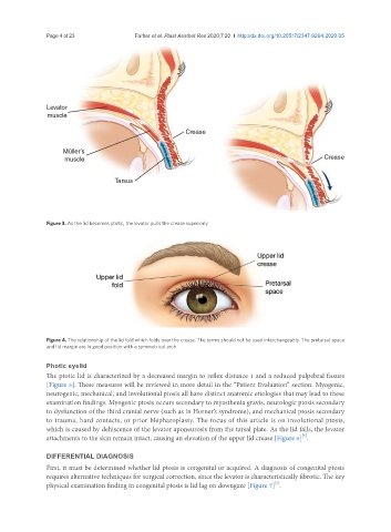
               Figure 3. As the lid becomes ptotic, the levator pulls the crease superiorly


               Figure 4. The relationship of the lid fold which folds over the crease. The terms should not be used interchangeably. The pretarsal space
               and lid margin are in good position with a symmetrical arch


               Photic eyelid
               The ptotic lid is characterized by a decreased margin to reflex distance 1 and a reduced palpebral fissure
               [Figure 5]. These measures will be reviewed in more detail in the “Patient Evaluation” section. Myogenic,
               neurogenic, mechanical, and involutional ptosis all have distinct anatomic etiologies that may lead to these
               examination findings. Myogenic ptosis occurs secondary to myasthenia gravis, neurologic ptosis secondary
               to dysfunction of the third cranial nerve (such as in Horner’s syndrome), and mechanical ptosis secondary
               to trauma, hard contacts, or prior blepharoplasty. The focus of this article is on involutional ptosis,
               which is caused by dehiscence of the levator aponeurosis from the tarsal plate. As the lid falls, the levator
                                                                                             [3]
               attachments to the skin remain intact, causing an elevation of the upper lid crease [Figure 6] .

               DIFFERENTIAL DIAGNOSIS
               First, it must be determined whether lid ptosis is congenital or acquired. A diagnosis of congenital ptosis
               requires alternative techniques for surgical correction, since the levator is characteristically fibrotic. The key
                                                                                     [4]
               physical examination finding in congenital ptosis is lid lag on downgaze [Figure 7] .

