
Page 208 - Read Online
P. 208
Farber et al. Plast Aesthet Res 2020;7:20 I http://dx.doi.org/10.20517/2347-9264.2020.05 Page 9 of 23
Figure 12. Bell’s test showing a good superior position of the eye on forced opening
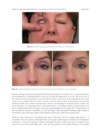
Figure 13. A patient with left upper lid ptosis with a high lid crease and elevation of the left brow for compensation
Next, the etiology of the patient’s lid ptosis should be determined as an extrinsic factor, such as brow ptosis
and pseudoptosis, or dermatochalasis, which can also give the appearance of a ptotic lid. In the case of
brow ptosis, the brow rests below its normal position, which is at the level of the supraorbital rim in men
or up to one centimeter above the rim in women. Attention must be paid to the presence of transverse
forehead rhytids and a unilateral elevated brow which is compensating for unilateral ptosis, as these may
be an indication of compensated brow or lid ptosis [Figure 13]. Dermatochalasis is characterized by an
[6]
excess of upper lid skin and soft tissue, which can cause pseudoptosis of the upper lid . In the presence of
true involutional lid ptosis, in addition to a low lid margin, other examination findings might include an
elevated lid crease and thinned upper eyelid, signifying attenuation of the levator aponeurosis .
[7]
There are many techniques for quantifying the degree of lid ptosis. First, the margin reflex distance 1 is
the distance from the central corneal light reflex to the upper eyelid margin. Normally, this measurement
is between 3-4 mm and is reduced in patients with upper lid ptosis. In the worst cases, the upper lid may
partially obstruct the corneal light reflex. Next the measurement of the palpebral fissure, or the eyelid