
Page 201 - Read Online
P. 201
Page 2 of 23 Farber et al. Plast Aesthet Res 2020;7:20 I http://dx.doi.org/10.20517/2347-9264.2020.05
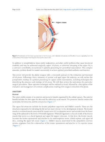
Figure 1. Attachments of the levator aponeurosis to the lid crease, which elevates with ptosis and Mueller’s muscle originating from the
undersurface of the levator inserting into the superior tarsal plate
In addition to xerophthalmia, lower eyelid malposition, and other eyelid problems that cause functional
disability and may be addressed surgically, upper lid ptosis, or abnormal drooping of the upper lid, is
a common comorbidity encountered in patients presenting for periorbital rejuvenation. This complex
anatomic problem should be treated concurrently to achieve optimal periorbital aesthetics and function.
This review will provide the plastic surgeon with a structured approach to the evaluation and treatment
of lid ptosis. Following a basic summary of normal and aged upper lid anatomy, we will outline the
preoperative workup of a patient presenting for upper eyelid rejuvenation, including techniques for
identifying the presence and etiology of lid ptosis. We will then review the indications for particular
surgical procedures. These operative techniques will be outlined in detail, followed by a description of the
evaluation and management of common complications resulting from surgical correction of lid ptosis.
ANATOMY
Normal
The upper eyelid consists of an anterior and posterior lamella separated by the orbital septum. The anterior
lamella includes the thin upper lid skin and the orbicularis oculi muscle. The posterior lamella contains the
[1]
tarsal plate, lid retractors, and the conjunctiva [Figure 1] .
The upper lid retractors include the levator palpebrae superioris and Müller’s muscle. These are the
structures responsible for elevating the lid and are most relevant to the development of ptosis. The levator
muscle is a striated muscle innervated by cranial nerve III (oculomotor nerve) and extends from the lesser
wing of the sphenoid to the level of Whitnall’s ligament. Whitnall’s ligament is a fascial sheath of the levator
muscle that serves as a check ligament and upper lid support structure. At this level, the levator muscle
becomes the levator aponeurosis and attaches to the anterosuperior tarsus, orbital septum, and upper lid
skin, creating the upper lid crease [Figure 2]. Müller’s muscle, innervated by the sympathetic nervous
system, originates from the undersurface of the levator aponeurosis and attaches to the superior tarsus
[1]
[Figure 3] .