
Page 58 - Read Online
P. 58
Walter et al. Unilateral rhinophyma
A B C
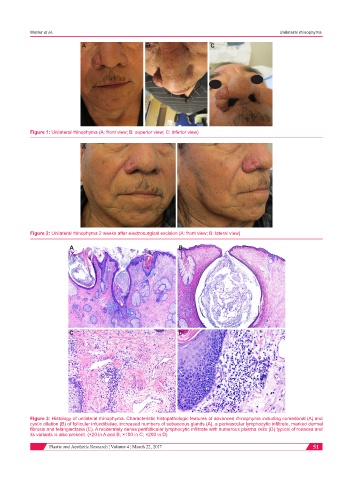
Figure 1: Unilateral rhinophyma (A: front view; B: superior view; C: inferior view)
A B
Figure 2: Unilateral rhinophyma 2 weeks after electrosurgical excision (A: front view; B: lateral view)
A B
C D
Figure 3: Histology of unilateral rhinophyma. Characteristic histopathologic features of advanced rhinophyma including comedonal (A) and
cystic dilation (B) of follicular infundibulae, increased numbers of sebaceous glands (A), a perivascular lymphocytic infiltrate, marked dermal
fibrosis and telangiectasia (C). A moderately dense perifollicular lymphocytic infiltrate with numerous plasma cells (D) typical of rosacea and
its variants is also present. (×20 in A and B; ×100 in C; ×200 in D)
Plastic and Aesthetic Research ¦ Volume 4 ¦ March 22, 2017 51