
Page 62 - Read Online
P. 62
Agrawal et al. Complications of Medpor® implants
A Outer skin envelope of nose with
columella everted
Fibrous covering of
Medpor implant incised
on one side
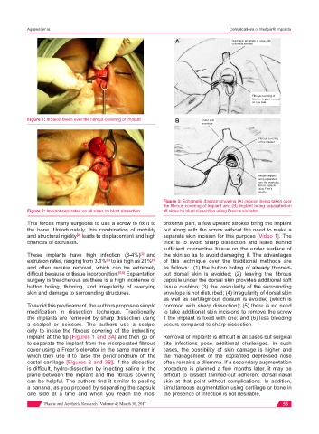
Figure 1: Incision taken over the fibrous covering of implant B Outer skin
envelope
Fibrous covering
of the implant
Medpor implant
being separated
from the overlying
fibrous capsule
using Free’s
elevator
Figure 3: Schematic diagram showing (A) incision being taken over
the fibrous covering of implant and (B) implant being separated on
Figure 2: Implant separated on all sides by blunt dissection all sides by blunt dissection using Freer’s elevator
This forces many surgeons to use a screw to fix it to proximal part, a few upward strokes bring the implant
the bone. Unfortunately, this combination of mobility out along with the screw without the need to make a
and structural rigidity leads to displacement and high separate skin incision for this purpose [Video 1]. The
[2]
chances of extrusion. trick is to avoid sharp dissection and leave behind
sufficient connective tissue on the under surface of
These implants have high infection (3-4%) [3] and the skin so as to avoid damaging it. The advantages
extrusion rates, ranging from 3.1% to as high as 21% of this technique over the traditional methods are
[4]
[5]
and often require removal, which can be extremely as follows: (1) the button holing of already thinned-
difficult because of tissue incorporation. [5,6] Explantation out dorsal skin is avoided; (2) leaving the fibrous
surgery is treacherous as there is a high incidence of capsule under the dorsal skin provides additional soft
button holing, thinning, and irregularity of overlying tissue cushion; (3) the vascularity of the surrounding
skin and damage to surrounding structures. envelope is not disturbed; (4) irregularity of dorsal skin
as well as cartilaginous dorsum is avoided (which is
To avoid this predicament, the authors propose a simple common with sharp dissection); (5) there is no need
modification in dissection technique. Traditionally, to take additional skin incisions to remove the screw
the implants are removed by sharp dissection using if the implant is fixed with one; and (6) less bleeding
a scalpel or scissors. The authors use a scalpel occurs compared to sharp dissection.
only to incise the fibrous covering of the indwelling
implant at the tip [Figures 1 and 3A] and then go on Removal of implants is difficult in all cases but surgical
to separate the implant from the incorporated fibrous site infections pose additional challenges. In such
cover using a Freer’s elevator in the same manner in cases, the possibility of skin damage is higher and
which they use it to raise the perichondrium off the the management of the explanted depressed nose
costal cartilage [Figures 2 and 3B]. If the dissection often remains a dilemma. If a secondary augmentation
is difficult, hydro-dissection by injecting saline in the procedure is planned a few months later, it may be
plane between the implant and the fibrous covering difficult to dissect thinned-out adherent dorsal nasal
can be helpful. The authors find it similar to peeling skin at that point without complications. In addition,
a banana, as you proceed by separating the capsule simultaneous augmentation using cartilage or bone in
one side at a time and when you reach the most the presence of infection is not desirable.
Plastic and Aesthetic Research ¦ Volume 4 ¦ March 30, 2017 55