
Page 57 - Read Online
P. 57
Walter et al. Unilateral rhinophyma
with a 1-year history of a growing mass on the right more frequent in skin phototypes I and II, though it is
nasal ala. The patient reported it started as a small increasingly being recognized as a condition seen in
papule and spread over the ala over the course of a all skin types. [2]
year; he noticed most of the growth during the last 4
months up to presentation. The patient denied any Although commonly diagnosed clinically, the differential
history of trauma to the area and denied manipulating diagnosis for rhinophyma should be considered,
the area. He denied using any topical medications especially when appearing unilaterally as in our
or products on his nose. He did not have a history of patient. Basal cell and squamous cell carcinomas can
similar lesions on the nose in the past. He denied any occur on phymatous skin, and should be considered
personal or family history of rosacea or of skin cancer. when unilateral changes, rapid growth, ulceration or
The patient was originally from El Salvador and then drainage occur. [2,3] Other neoplasms including adnexal
immigrated to the United States. He was retired from tumors would also be included and can be considered.
his work at the time of presentation. He previously Granulomatous processes such as sarcoidosis and
worked outdoors in construction for many years and infectious diseases such as rhinoscleroma (Klebsiella)
had a number of sunburns in the past. or leishmania should also be considered in the
appropriate clinical setting.
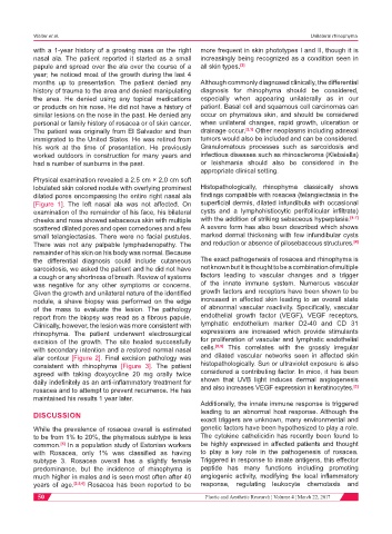
Physical examination revealed a 2.5 cm × 2.0 cm soft
lobulated skin colored nodule with overlying prominent Histopathologically, rhinophyma classically shows
dilated pores encompassing the entire right nasal ala findings compatible with rosacea (telangiectasia in the
[Figure 1]. The left nasal ala was not affected. On superficial dermis, dilated infundibula with occasional
examination of the remainder of his face, his bilateral cysts and a lymphohistiocytic perifollicular infiltrate)
cheeks and nose showed sebaceous skin with multiple with the addition of striking sebaceous hyperplasia. [4-7]
scattered dilated pores and open comedones and a few A severe form has also been described which shows
small telangiectasias. There were no facial pustules. marked dermal thickening with few infundibular cysts
There was not any palpable lymphadenopathy. The and reduction or absence of pilosebaceous structures. [4]
remainder of his skin on his body was normal. Because
the differential diagnosis could include cutaneous The exact pathogenesis of rosacea and rhinophyma is
sarcoidosis, we asked the patient and he did not have not known but it is thought to be a combination of multiple
a cough or any shortness of breath. Review of systems factors leading to vascular changes and a trigger
was negative for any other symptoms or concerns. of the innate immune system. Numerous vascular
Given the growth and unilateral nature of the identified growth factors and receptors have been shown to be
nodule, a shave biopsy was performed on the edge increased in affected skin leading to an overall state
of the mass to evaluate the lesion. The pathology of abnormal vascular reactivity. Specifically, vascular
report from the biopsy was read as a fibrous papule. endothelial growth factor (VEGF), VEGF receptors,
Clinically, however, the lesion was more consistent with lymphatic endothelium marker D2-40 and CD 31
rhinophyma. The patient underwent electrosurgical expressions are increased which provide stimulants
excision of the growth. The site healed successfully for proliferation of vascular and lymphatic endothelial
with secondary intention and a restored normal nasal cells. [8,9] This correlates with the grossly irregular
alar contour [Figure 2]. Final excision pathology was and dilated vascular networks seen in affected skin
consistent with rhinophyma [Figure 3]. The patient histopathologically. Sun or ultraviolet exposure is also
agreed with taking doxycycline 20 mg orally twice considered a contributing factor. In mice, it has been
daily indefinitely as an anti-inflammatory treatment for shown that UVB light induces dermal angiogenesis
[3]
rosacea and to attempt to prevent recurrence. He has and also increases VEGF expression in keratinocytes.
maintained his results 1 year later.
Additionally, the innate immune response is triggered
leading to an abnormal host response. Although the
DISCUSSION
exact triggers are unknown, many environmental and
While the prevalence of rosacea overall is estimated genetic factors have been hypothesized to play a role.
to be from 1% to 20%, the phymatous subtype is less The cytokine cathelicidin has recently been found to
common. In a population study of Estonian workers be highly expressed in affected patients and thought
[5]
with Rosacea, only 1% was classified as having to play a key role in the pathogenesis of rosacea.
subtype 3. Rosacea overall has a slightly female Triggered in response to innate antigens, this effector
predominance, but the incidence of rhinophyma is peptide has many functions including promoting
much higher in males and is seen most often after 40 angiogenic activity, modifying the local inflammatory
years of age. [3,5,6] Rosacea has been reported to be response, regulating leukocyte chemotaxis and
50 Plastic and Aesthetic Research ¦ Volume 4 ¦ March 22, 2017