
Page 183 - Read Online
P. 183
Bhandari et al. Metacarpal angulations
Table 1: Dorsal cortical angle Table 2: Center of rotation of angulation
Mean (°) Range (°) Standard deviations Mean (%) Range (%) Standard deviations
2nd 13 6-26 4.73 2nd 53.5 42-85 12.5
3rd 10 1-25 5.28 3rd 52.1 32-100 17.8
4th 11 1-20 4.45 4th 48.3 34-86 11.5
5th 12 2-24 5.11 5th 50.3 27-86 12.4
(range, 2-24°; SD, 5.11). There was no progressive bony eminences in case of a concave bony ending,
difference as we moved from the II digit to the V digit. as a line through the points indicating the widest
osseous space in case of a convex cortex ending, and
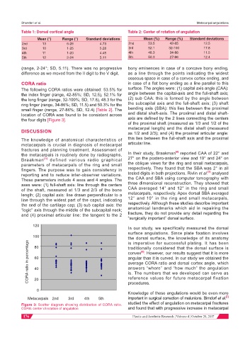
CORA ratio in case of a flat bony ending as a line parallel to this
The following CORA ratios were obtained: 53.5% for surface. The angles were: (1) capital axis angle (CAA):
the index finger (range, 42-85%; SD, 12.5), 52.1% for angle between the capital-axis and the full-shaft axis;
the long finger (range, 32-100%; SD, 17.8), 48.3 for the (2) sub CAA: this is formed by the angle between
ring finger (range, 34-86%; SD, 11.5) and 50.3% for the the subcapital axis and the full-shaft axis; (3) shaft
small finger (range, 27-86%; SD, 12.4) [Table 2]. The bending axis (SBA): this lies between the proximal
location of CORA was found to be consistent across and distal shaft-axis. The proximal and distal shaft-
the four digits [Figure 3]. axis are defined by the 2 lines connecting the centers
of the proximal shaft (measured as 1/3 and 1/2 of the
metacarpal length) and the distal shaft (measured
DISCUSSION as 1/2 and 2/3); and (4) the proximal articular angle:
The knowledge of anatomical characteristics of this lies between the full-shaft axis and the proximal
metacarpals is crucial in diagnosis of metacarpal articular line.
fractures and planning treatment. Assessment of [4]
the metacarpals is routinely done by radiographs. In their study, Braakman reported CAA of 22° and
Braakman [4] defined various radio graphical 27° on the postero-anterior view and 19° and 24° on
parameters of metacarpals of the ring and small the oblique views for the ring and small metacarpals,
fingers. The purpose was to gain consistency in respectively. They found that the SBA was 2° in all
[5]
reporting and to reduce inter-observer variations. tested digits in both projections. Rivlin et al. analysed
These parameters include 4 axes and 4 angles. The the CAA and SBA using computer tomography with
axes were: (1) full-shaft axis: line through the centers three dimensional reconstruction. They showed that
of the shaft, measured at 1/3 and 2/3 of the bone CAA averaged 14° and 12° in the ring and small
length; (2) capital axis: line drawn perpendicular to a metacarpals, respectively. Apex dorsal SBA averaged
line through the widest part of the caput, indicating 12° and 10° in the ring and small metacarpals,
the end of the cartilage cap; (3) sub capital axis: the respectively. Although these studies describe important
“logic” axis through the middle of the subcapital neck; anatomical landmarks which aid in repairing the
and (4) proximal articular line: the tangent to the 2 fracture, they do not provide any detail regarding the
“surgically important” dorsal surface.
120
In our study, we specifically measured the dorsal
surface angulations. Since plate fixation involves
100 the dorsal surface, the knowledge of its anatomy
CORA ratio in percentage 60 convex . However, our results suggest that it is more
is imperative for successful plating. It has been
80
traditionally considered that the dorsal surface is
[6]
angular than it is curved. In our study we obtained the
average CORA ratio and dorsal cortex angle, which
answers “where” and “how much” the angulation
40
is. The numbers that we developed can serve as
20
procedures.
0 reference values for future metacarpal fixation
Knowledge of these angulations would be even more
[7]
Metacarpals 2nd 3rd 4th 5th important in surgical correction of malunions. Birndorf et al.
studied the effect of angulation on metacarpal fractures
Figure 3: Scatter diagram showing distribution of CORA ratio.
CORA: center of rotation of angulation and found that with progressive increase in metacarpal
176 Plastic and Aesthetic Research ¦ Volume 4 ¦ October 20, 2017