
Page 98 - Read Online
P. 98
of the genital and anorectal region, and in tumors healing. Primary reconstruction of the groin should,
involving the lower extremities. The clinical presentation therefore, always be considered for patients undergoing
[1]
of locally advanced primary and nodal disease is ilio-inguinal node dissection.
not uncommon in India. Surgery may be curative or Trauma to the inguinal region with soft tissue defects is not
palliative in such presentations, which requires radical uncommon. A high index of suspicion for injuries to the
surgery for the primary tumor and en bloc inguinal or femoral vessels is needed in such cases. With soft tissue
ilio-inguinal lymphadenectomy. Inguinal node dissection defects over the inguinal region, there is always a need for
has been always associated with a high incidence of stable soft tissue coverage over the femoral vessels.
wound complications. The surgical oncologist has
moved from radical dissection to sentinel lymph node Aims of primary reconstruction of the soft tissue defects
dissection to reduce the morbidity due to surgery. Still over the groin region are protection of the femoral
the role of radical inguinal lymphadenectomy cannot be vessels, provision of well-vascularized tissue from a distant
avoided in certain situations. Potential complications area, coverage of the dead space in the femoral triangle,
following inguinal block dissection are infection (6–20%), a decrease in seroma formation, wound closure without
lymphorrhea (6–40%), lymphedema (8–69%) and skin flap tension, initiation of radiotherapy as early as possible,
necrosis (27–85%). Removal of the adipofascial layer and a decrease in the length of the hospital stay. [3]
[2]
in a groin dissection damages the subdermal plexus, Reconstructive options available for coverage of inguinal
potentially leading to skin flap necrosis. To reduce defects include the random pattern flap, the tensorfascia lata
complications-related to wound healing, various primary flap, the perforator propeller-type TFL flap, the modified TFL
reconstructive procedures such as muscle transposition flap, the gracilis and sartorius flaps, the anterolateral thigh
and myocutaneous flaps are used for groin reconstruction. flap, the omental flap, the rectus femoris flap and the rectus
Many of these patients require adjuvant radiotherapy abdominis flap. Skin grafting is not sufficient for stable
following surgery. Hence, these patients require stable coverage over exposed bones, nerves and vessels. Free
skin coverage over the operated site for the prevention tissue transfer requires enhanced microsurgical expertise
of tissue edema, fibrosis and complications due to wound and may overburden patients in critical condition with
progressive malignant disease. In such situations, sufficient
soft tissue coverage can be achieved by simple and reliable
techniques with minimal donor site morbidity.
The TFL flap was first described in 1934 by Wangensteen
[4]
and was popularized by Nahai et al. for the reconstruction
[5]
of pressure ulcer defects and for complications following
block dissections. Disadvantages of the TFL flap include
a b
proximal bulkiness with a thin distal flap, a depressed
donor region with an unsightly appearance of the grafted
area, and potential loss of stability of the knee due to the
sacrifice of fascia lata.
The modified TFL flap includes the muscle with a hatchet
shaped incision, which provides adequate mobility of the
flap and reduces the dog ear deformity, ensuring closure
c
of the donor area without the need for a skin graft or
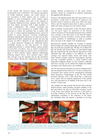
Figure 3: The patient with fungating right sided inguinal lymph nodes. local flap. Variations in the incision for the flap may not
(a) Soft tissue defect inguinal region following dissection; (b) planning of significantly contribute to the reduction of the donor
superiorly-based perforator plus flap; (c) immediate postoperative view
of the superiorly-based perforator plus flap site deformity when the muscle is included in the flap.
a b c
d e f g
Figure 4: The patient with soft tissue defect over the inguinal region. (a) Soft tissue defect right inguinal region following trauma; (b) closer view
of contaminated soft tissue defect groin region; (c) soft tissue defect following debridement; (d) planning of superiorly-based perforator plus flap;
(e) identification and preserving the perforators entering the flap; (f) immediate postoperative view of the superiorly-based perforator plus flap; (g) late
postoperative picture
92 Plast Aesthet Res || Vol 1 || Issue 3 || Dec 2014