
Page 97 - Read Online
P. 97
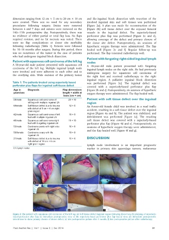
dimension ranging from 12 cm × 5 cm to 20 cm × 10 cm   and ilio-inguinal block dissection with resection of the
          were created. There was no need for any secondary   involved inguinal skin and soft tissues was performed
          procedures following surgery. Drains were removed   [Figure 2a]. A plan was made for reconstruction of the
          between 4 and 7 days and sutures were removed on the   [Figure 2b] soft tissue defect over the exposed femoral
          14th–17th postoperative day. Postoperatively, there was   vessels in the inguinal defect. The superiorly-based
          no evidence of either partial or total flap loss. No flaps   perforator plus flap was performed [Figure 2c and d],
          required revision, and no fat necrosis was noted. There   allowing coverage of the defect and primary closure of
          were no flap complications or donor site morbidity   the donor site defect. Postoperatively, six sessions of
          following radiotherapy [Table 1]. Patients were followed   hyperbaric oxygen therapy were administered. The flap
          for 10–18 months after surgery. During this period, there   healed well [Figure 2e and f]. Regular follow-up was
          was no recurrence of the tumor in the case of patients   performed. The flap tolerated radiotherapy well.
          who had undergone inguinal block dissection.
                                                              Patient with fungating right-sided inguinal lymph
          Patient with squamous cell carcinoma of the left leg  nodes
          A 58-year-old male patient presented with squamous cell   A 48-year-old male patient presented with fungating
          carcinoma of the left leg. Multiple inguinal lymph node   inguinal lymph nodes on the right side. He had previously
          were involved and were adherent to each other and to   undergone surgery for squamous cell carcinoma of
          the overlying skin. Wide excision of the primary lesion
                                                              the right foot and received radiotherapy to the right
                                                              inguinal region. A palliative inguinal block dissection
          Table 1: The patients treated using superiorly based   was performed [Figure 3a]. The inguinal defect was
          perforator plus fl aps for inguinal soft tissue defect  covered with a superiorly-based perforator plus flap
           Age in    Diagnosis               Flap dimensions   [Figure 3b and c]. Postoperatively, six sessions of hyperbaric
           years/sex                         length × width at   oxygen therapy were administered. The flap healed well.
                                              base (cm × cm)
           59/male   Squamous cell carcinoma of   20 × 10     Patient with soft tissue defect over the inguinal
                     left leg with multiple inguinal LN       region
           8/female  Soft tissue defect due to trauma   12 × 5  An 8-year-old female child was involved in a road traffic
                     with defect of 9 cm × 4 cm right         accident, resulting in a soft tissue defect over the inguinal
                     groin region
           42/male   Squamous cell carcinoma of left   18 × 9  region [Figure 4a and b]. The patient was stabilized, and
                     foot with multiple inguinal LN           debridement was performed [Figure 4c]. The resulting
           48/male   Squamous cell carcinoma right   18 × 10  soft tissue defect was covered with a superiorly-based
                     foot with fungating inguinal LN          perforator plus flap [Figure 4d and e]. Postoperatively, six
           52/male   Carcinoma penis with right side   16 × 8  sessions of hyperbaric oxygen therapy were administered,
                     inguinal LN                              and the flap healed well [Figure 4f and g].
           56/female  Carcinoma ovary with ilio   19 × 9
                     inguinal LN
           27/male   Soft tissue defect due to trauma   15 × 6  DISCUSSION
                     with defect of 14 cm × 6 cm
                     right groin region                       Lymph node involvement is an important prognostic
           LN: Lymph nodes                                    marker in primary skin appendage tumors, melanomas


                         a                   b                         c


                         d                       e                        f
          Figure 2: The patient with squamous cell carcinoma of the left leg. (a) Soft tissue defect inguinal region following dissection; (b) planning of superiorly-
          based perforator plus flap; (c) immediate postoperative view of the superiorly-based perforator plus flap lateral view; (d) immediate postoperative
          lateral view to show primary closure of donor defect; (e) late postoperative picture after 1 month; (f) late postoperative picture after radiotherapy

          Plast Aesthet Res || Vol 1 || Issue 3 || Dec 2014                                                 91