
Page 101 - Read Online
P. 101
then placed following a modified drilling sequence to
undersize the osteotomy and increase the insertion
torque. The implant system’s drills were of kind implant
fixture with parallel shape. Drills set a low speed were
used in succession, harvesting autogenous bone from the
drills for later bone defect grafting. A precision initial drill
allowed accurate positioning of the osteotomy within the
palatal alveolar wall. Once the direction of drilling was
established, the site was enlarged with a 2 mm pilot drill.
Subsequent twist drills were used to widen the osteotomy
following the manufacturer’s instructions. The final drill
however, was only utilized to a depth of approximately
two-thirds of the implant length.
Implant site was prepared using a surgical motor
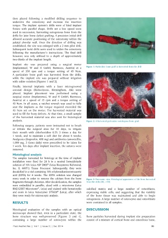
(Implantmed, W and H GmbH, Burmoos, Austria) at a Figure 1: Particulate bone graft is harvested from the drill
speed of 350 rpm and a torque setting of 45 Ncm.
A particulate bone graft was harvested from the drills,
while the implant site was prepared without irrigation
with saline solution [Figures 1 and 2].
Finally, internal implants with a laser microgrooved
coronal design (Biohorizons, Birmingham, Ala) were
placed. Implant placement was performed using a
surgical motor (Implantmed, W and H GmbH, Burmoos,
Austria) at a speed of 15 rpm and a torque setting of
45 Ncm. In all cases, a ratchet wrench was used to fully
seat the implants as the torque required exceeded the
45 Ncm set on the motor. The harvested material was
used to fill the bony defects. At that time, a small sample
of the harvested material was also sent for histological
analysis. Figure 2: A harvested particulate autologous bone graft
Following surgery, patients were instructed not to brush
or irritate the surgical sites for 10 days, to irrigate
their mouth with chlorhexidine 0.2% 3 times a day for
1 week, and to maintain a soft diet for about 6 weeks.
Analgesics (ibuprofen, 400 mg) and antibiotics (amoxicillin,
1,000 mg, 3 times daily) were prescribed to be taken for
1 week. Ten days after implant insertion, the sutures were
removed.
Histological analysis
The samples harvested for histology at the time of implant
installation were fixed for 24 h in a neutral formaldehyde
solution of 10% Leica ASP 300S (Leica Biosystems Richmond,
®
Inc. IL 60071) Tissue Processor. Subsequently, they were
decalcified in a vial containing 10% ethylenediaminetetraacetic
acid (EDTA) for 4 weeks. The EDTA solution was changed
every week in order to remove the calcium from the bone Figure 3: Panoramic view. Histological appearance of the bone harvested
fragments through chelation. After decalcification, the samples from the drills (HE, ×10)
were embedded in paraffin, sliced with a microtome (Leica
RM2125RT Microtome , Leica) and stained with hematoxylin calcified matrix and a large number of osteoblasts,
®
and eosin in Leica Autostainer ST5020 (Leica), after which expressing viable cells, and suggesting that the viability
®
they were ready for microscopic analysis. of the bone tissue was maintained and able to begin
ostogenesis. A large number of osteocytes and osteoblasts
RESULTS were contained in all samples.
Histological evaluation of the samples with an optical DISCUSSION
microscope showed that, even in a particulate state, the
bone structure was well-preserved [Figures 3 and 4], Bone particles harvested during implant site preparation
containing a large number of osteocytes within the consist of a mixture of cortical bone and cancellous bone,
Plast Aesthet Res || Vol 1 || Issue 3 || Dec 2014 95