Page 93 - Read Online
P. 93

DISCUSSION                                          to the following formula: P = 2R × 3.14 = d × 3.14 (R:
                                                              radius = d/2). This measurement allowed us to determine
          Either the DGA or SGA perfuse the medial femoral condyle.   the outer diameter in noninflated arteries.
          The current study evaluated the anatomical structures and   Our study detected the DGA in 82.5% and the
          variations of the DGA and SGA as well as their branches in   SGA in 25% of the 40 specimens. Yamamoto
          adult cadavers. One half of cadavers were studied in fresh   et al.  found the DGA in 89% and the SGA in 100% of the
                                                                  [7]
          condition to facilitate the evaluation of the perfusion area   19 specimens. Rahmanian-Schwarz  et  al.  harvested the
                                                                                                 [1]
          of skin and bone.
                                                              DGA in 100% of the 21 specimens and Iorio  et  al.  also
                                                                                                         [16]

          Prior studies have shown that the outer diameter of   discovered the DGA in 100% of the 12 specimens. The
          the artery from fresh frozen cadavers was maintained.   difference between the studies is secondary to the number
          While the diameter of arteries from cadavers preserved   of specimens.
          in formalin retracts and loses its shape, the perimeter of   In the current study, the dominant vessels supplying the
          the artery is maintained.  We calculated the outer   medial femoral condyle were the DGA in 82.5% of cases
                                 [7]
          diameter of the artery through its perimeter  according
                                                              and the SGA in the DGA and 17.5% of cases. Van Dijck
                                                              et al.  showed that in 70% of cases the DGA was dominant,
                                                                  [6]
                                                              while in 21% of cases the SGA was the dominant vessel. In
                                                              9% of cases the DGA and SGA supplied the medial femoral
                                                              condyle equally. A comparison between the measurements
                                                              of the DGA in the current study and other studies is made
                                                              in Table 1.
                                                              Similar to previous studies, the current study
                                                              demonstrated that the DGA generally divides into 3
                                                              branches (63.7% of cases) or 2 branches (33.3% of cases).
                                                              In addition, the AB of the DGA or the SGA always
                                                              nourishes the periosteum of the medial femoral condyle.
                                                              These arteries have adequate diameter and length to
                                                              supply the medial femoral condylar flap. In the absence
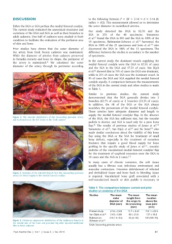
          Figure 1:  The vascular distribution of the descending genicular artery   of the DGA, the SGA has sufficient size, but the vascular
          and its branches at the left femur in the fresh cadaver
                                                              pedicle is shorter, and SGA is used only for a pure bone
                                                                  [8]
                                                              flap.  The results of previous studies of Jones  et al.
                                                                                                             [10]
                                                                                            [6]
                                                                                                         [13]
                                                              Yamamoto  et al. , Van Dijck  et al.  and De Smet  also
                                                                            [7]
                                                              made similar conclusions about the viability of this bone
                                                              flap using the DGA or the SGA for treatment of small
                                                              bony defects, especially in the treatment of nonunion
                                                              fractures that require a good blood supply for bone
                                                              grafting. In the specific study of Jones  et  al. , vascular
                                                                                                     [11]

                                                              pedicles of the vascularized medial femoral condylar flap
                                                              for the treatment of scaphoid nonunion were the DGA in
                                                              10 cases and the SGA in 2 cases. [11]
                                                              In many cases of chronic nonunion, the soft tissue
                                                              usually has a fibrous scar, infectious environment and
                                                              avascular contracture. Extensive debridement of infected
          Figure 2: Anatomy of the articular branch from the descending genicular   and devitalized tissue and bone back to bleeding tissue
          artery for blood supply to the medial femoral condyle  is required. Vascularized bone graft associated with a
                                                              well-vascularized muscle or skin paddle is necessary in

                                                              Table 1: The comparison between current and prior
                                                              studies on anatomy of the DGA
                                                               Studies       The mean     The mean    The mean
                                                                               outer     length from   location
                                                                             diameter of   the origin to   above the
                                                                             DGA (mm)   its branching   knee joint
                                                                                           (mm)         (mm)
                                                               Present study  2.16 ± 0.69  11.7 ± 8.61  119.1 ± 23.6
                                                               Van Dijck et al. [6]  2.43 ± 0.88  89 ± 21.8  137 ± 18.8
                                                               Rahmanian-    2.9 (1.5–4.5)  25 (5–40)  147 (120–70)
          Figure 3: Cutaneous angiosome distribution of the saphenous branch at   Schwarz et al. [1]
          the medial side of the knee and proximal leg after injected methylene
          blue in fresh cadavers                               DGA: Descending genicular artery
          Plast Aesthet Res || Vol 1 || Issue 3 || Dec 2014                                                 87
   88   89   90   91   92   93   94   95   96   97   98